
Fig. 6
- ID
- ZDB-IMAGE-090109-17
- Publication
- Linville et al., 2009 - Combinatorial roles for zebrafish retinoic acid receptors in the hindbrain, limbs and pharyngeal arches
- All Figures
- Figures for Linville et al., 2009
|
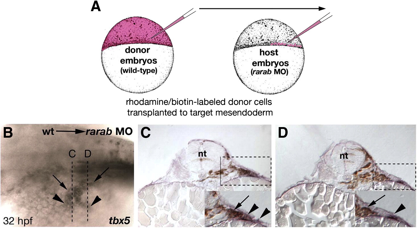
Fig. 6 Grafts of wild-type mesoderm rescue tbx5 expression in rarab morphants. (A) Cell transplantation technique. Donor embryos were injected at the one-cell stage with both a fixable biotinylated dextran and a fluorescent rhodamine-dextran (red). Hosts were simultaneously injected with the rarab MO. At 4 hpf small groups of cells were transferred from donors to the margin (which forms the mesendoderm) of hosts, which were raised to 32 hpf, and stained for biotin (brown, grafted cells) and tbx5 (blue, pectoral fin). (B) Whole mount, anterior to the left, showing partial unilateral rescue of tbx5 expression — arrowheads indicate tbx5-expressing cells, and arrows indicate locations of wild-type, donor cells. Rescue was either partial (15/50) or completely restored the fin (2/50). Dotted lines indicate locations of transverse sections in panels C and D. (C, D) Transverse sections through the fin fields of mosaic embryos, showing the absence of tbx5 expression on the untransplanted, left side and tbx5+ cells in the LPM (arrowheads) on the right side in close proximity to biotin-labeled, donor cells (arrows). Abbreviations: nt, neural tube; wt, wild type.
Reprinted from Developmental Biology, 325(1), Linville, A., Radtke, K., Waxman, J.S., Yelon, D., and Schilling, T.F., Combinatorial roles for zebrafish retinoic acid receptors in the hindbrain, limbs and pharyngeal arches, 60-70, Copyright (2009) with permission from Elsevier. Full text @ Dev. Biol.