
Image
|
Figure Caption
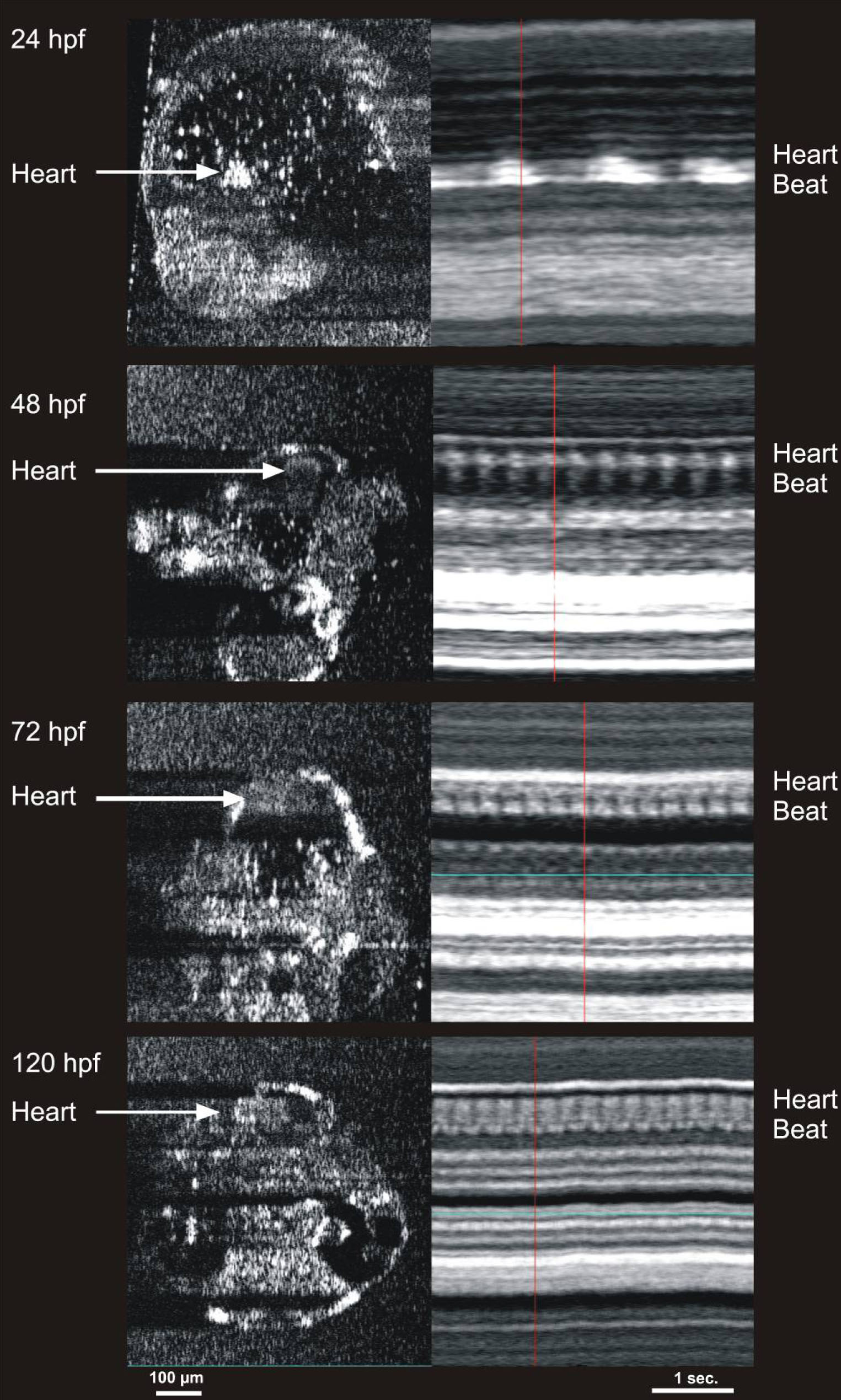
Fig. 8 Cardiac M-mode images of the heart in 24, 48, 72, and 120 hours post fertilization embryos. The bright signal was created by blood within the heart. Note the increase in heart rate with development, as well as the development of two chambers at 72 hours post fertilization (hpf). The heart rates observed in the m-mode images are 47 beats per min (bpm) in the 24 hpf embryo, 157 bpm in the 48 hpf embryo, 219 bpm in the 72 hpf embryo, and 250 bpm in the 120 hpf embryo.
Acknowledgments
This image is the copyrighted work of the attributed author or publisher, and
ZFIN has permission only to display this image to its users.
Additional permissions should be obtained from the applicable author or publisher of the image.