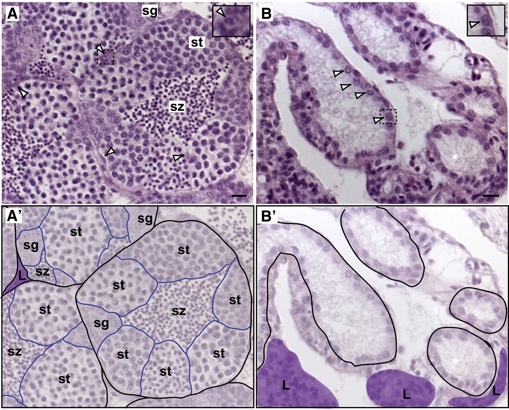
Fig. 1 Zebrafish lacking germ cells make testes. H&E stainings of adult (7 month old) wild-type and germ line deficient testes. (A) Wild-type testis. The testis is organized in tubules surrounded by basement membrane, outlined in black (A′). Within the tubules, cysts of germ cells undergo spermatogenesis synchronously, outlined in blue (A′). Spermatozoa (sz) are released into the center, which connects to the efferent ducts. Sertoli cells (arrowheads and inset) are found within the tubules and Leydig cells (L) are in the interstitial spaces, colored purple (A′). (B) Germ line deficient testis. Testis tubules are formed, outlined in black (B′). Sertoli cells are lining the inside of the tubules (some marked by arrowheads and shown in inset). Leydig cells (L) are intertubular, colored purple (B′). Vasculature is also present in intertubular regions. sz, spermatozoa; st, spermatids; sg, spermatogonia. Scale bars are 10 μm.
Reprinted from Developmental Biology, 324(2), Siegfried, K.R., and Nüsslein-Volhard, C., Germ line control of female sex determination in zebrafish, 277-287, Copyright (2008) with permission from Elsevier. Full text @ Dev. Biol.

