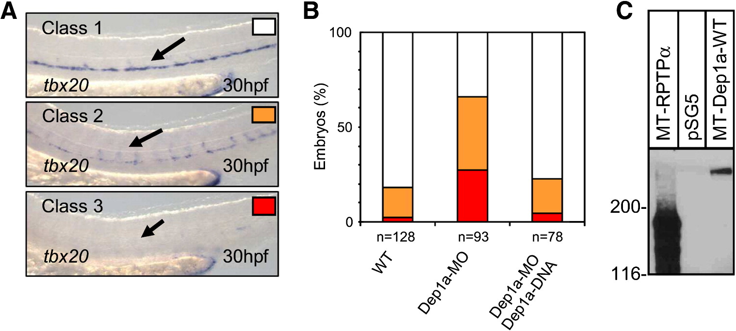
Fig. 5 Dep1a-MO-induced reduction in tbx20 expression was specific. (A) Classification of the observed defects after Dep1a-MO injection (8 ng/embryo): upper panel, no effect, normal tbx20 expression (white), middle panel, mild effect, patchy tbx20 expression (orange), lower panel, severe effect, no tbx20 expression (red). (B) Quantification of the effects of Dep1a knockdown. The percentages of embryos in the three different classes is given in control embryos (WT) and in Dep1a-MO1 (8 ng) injected embryos. Moreover, Dep1a-MO1 was co-injected with a CMV promoter-driven expression vector for Dep1a (12.5 ng), effectively rescuing the defects. Total numbers of embryos from at least three independent experiments are indicated (n). Exact numbers are given in Supplementary Table S1. (C) Expression vectors for Myc epitope tagged Dep1a and – as a control – for RPTPα were transfected into COS cells. The cells were lysed and whole cell lysates were run on a 7.5% SDS-PAAGE gel. The material on the gel was blotted and the blots were probed with anti-Myc MAb 9E10. An immunoblot is depicted developed with enhanced chemiluminescence and the positions of marker proteins that were co-electrophoresed with the sample are indicated in kDa on the left.
Reprinted from Developmental Biology, 324(1), Rodriguez, F., Vacaru, A., Overvoorde, J., and den Hertog, J., The receptor protein-tyrosine phosphatase, Dep1, acts in arterial/venous cell fate decisions in zebrafish development, 122-130, Copyright (2008) with permission from Elsevier. Full text @ Dev. Biol.

