
Image
|
Figure Caption
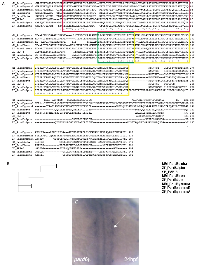
Fig. S4 The Pard6 protein family. (A) Clustal W (1.83) Multiple Sequence Alignment of the four zebrafish (ZF) Pard6 proteins with the three mouse (MM) Pard6 proteins and the C.elegans (CE) PAR-6 protein. The red box outlines the conserved PB1 domain. The green box outlines the conserved CRIB domain. The yellow boxes outline the conserved PDZ domain. (B) Cladogram of the four zebrafish (ZF) Pard6 proteins, three mouse (MM) Pard6 proteins, and the C.elegans (CE) PAR-6 proteins. (C) in situ hybridization analysis shows pard6β expression at 24 hpf in the otic placodes (arrow).
Figure Data
Acknowledgments
This image is the copyrighted work of the attributed author or publisher, and
ZFIN has permission only to display this image to its users.
Additional permissions should be obtained from the applicable author or publisher of the image.
Reprinted from Developmental Biology, 324(1), Munson, C., Huisken, J., Bit-Avragim, N., Kuo, T., Dong, P.D., Ober, E.A., Verkade, H., Abdelilah-Seyfried, S., and Stainier, D.Y., Regulation of neurocoel morphogenesis by Pard6gammab, 41-54, Copyright (2008) with permission from Elsevier. Full text @ Dev. Biol.