
|
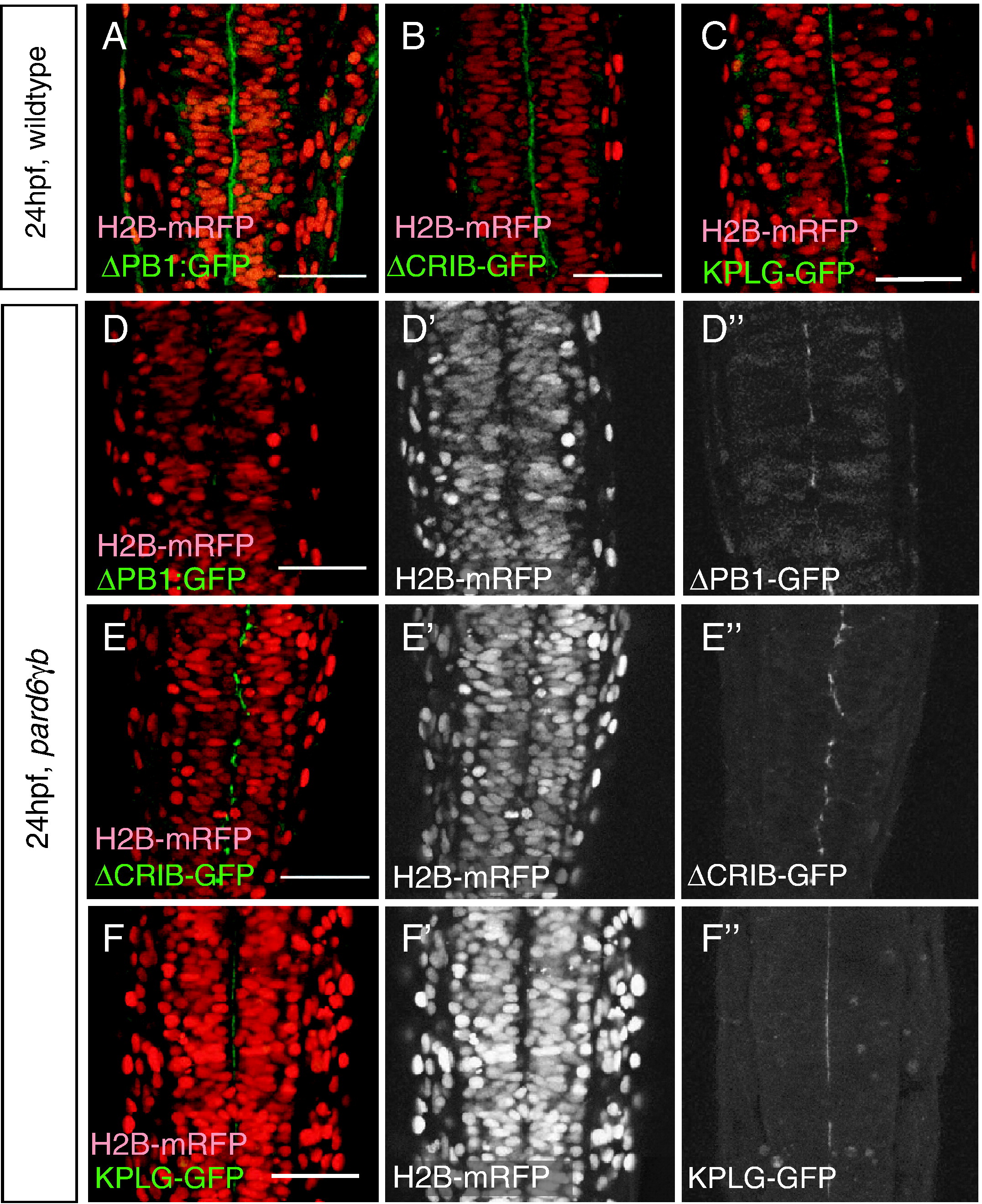
Fig. 6 The PB1, CRIB and PDZ domains are required for Pard6γb function but not localization. (A–E) Live, 24 hpf embryos injected with h2b-mRFP and pard6γb(Δpb1)-GFP (abbreviated ΔPB1-GFP), pard6γb(Δcrib)-GFP (abbreviated ΔCRIB-GFP) or pard6γb(kplg)-GFP (abbreviated KPLG-GFP) mRNA as indicated (dorsal views between the first and sixth somite, anterior to the top). (A–C) In wildtype neural tubes, nuclei are organized on either side of the midline. (D-F) In pard6γbs441 mutant neural tubes, nuclei are disorganized. In wildtype embryos, ΔPB1-GFP (A), ΔCRIB-GFP (B) and KPLG-GFP (C) localized to the cytoplasm and apical membranes. In pard6γbs441 mutant embryos, ΔPB1-GFP (D), ΔCRIB-GFP (E) and KPLG-GFP (F) failed to rescue the mutant phenotype and localized to the discontinuous apical membranes. Scale bars represent 50 μm.
Reprinted from Developmental Biology, 324(1), Munson, C., Huisken, J., Bit-Avragim, N., Kuo, T., Dong, P.D., Ober, E.A., Verkade, H., Abdelilah-Seyfried, S., and Stainier, D.Y., Regulation of neurocoel morphogenesis by Pard6gammab, 41-54, Copyright (2008) with permission from Elsevier. Full text @ Dev. Biol.