
Fig. 5
- ID
- ZDB-IMAGE-081202-11
- Publication
- Munson et al., 2008 - Regulation of neurocoel morphogenesis by Pard6gammab
- All Figures
- Figures for Munson et al., 2008
|
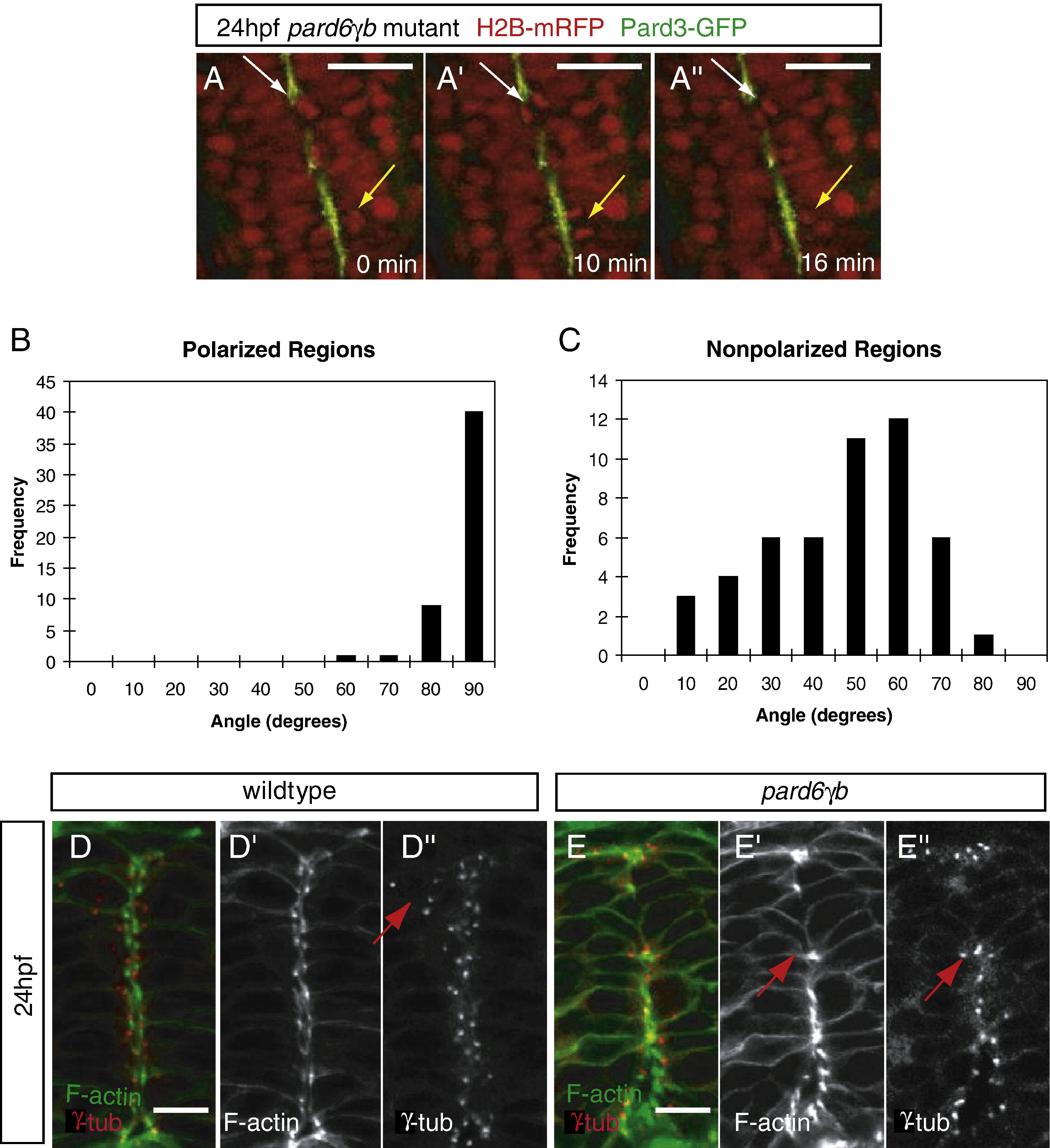
Fig. 5 Position of the apical membranes in the neural tube appears to regulate the orientation of mitoses and the positioning of centrosomes during the neural tube stage. (A) Time lapse images of a 24 hpf pard6γbs441 mutant embryo injected with pard3-gfp and h2b-mrfp mRNA (dorsal views between the first and sixth somite, anterior to the top). Time is indicated in minutes. The yellow arrow points to a cell division in a region of continuous polarity, at an 88° angle (see B). The white arrow points to a cell division in a region of discontinuous polarity, at a 26° angle (see panel C). (B, C) Schematic of the technique used to analyze the angle of cell division in wildtype and pard6γbs441 mutant embryos. The angle between the midline (green) of the neural tube and the cleavage plane (black) of two dividing nuclei was recorded and analyzed on a histogram using the value ≤ 90°. (B) In regions with polarized localization of Pard3-GFP in wildtype (n = 3) and pard6γbs441 mutant (n = 12) embryos, the angle of division clustered between 80° and 90° (n = 40/50). (C) In regions lacking polarized localization of Pard3-GFP in pard6γbs441 mutant embryos, the angle of division clustered between 40° and 70° (n = 29/50). (D, E) Transverse sections through the neural tube between the first and sixth somite of 24 hpf embryos, stained for filamentous actin and γ-Tubulin. (D) In wildtype embryos, centrosomes are localized along the apical side of the cells lining the neurocoel or rotated 90° when cells are dividing (arrow). (panel F) In pard6γbs441 mutant embryos, centrosomes are positioned as in wildtype when polarized apical membranes are present, but appear to be missing or mislocalized in nonpolarized regions. The arrow points to a cell that has re-oriented the position of its centrosome towards a polarized region. Scale bars in panel A represent 50 μm, and in panels D and E 10 μm.
Reprinted from Developmental Biology, 324(1), Munson, C., Huisken, J., Bit-Avragim, N., Kuo, T., Dong, P.D., Ober, E.A., Verkade, H., Abdelilah-Seyfried, S., and Stainier, D.Y., Regulation of neurocoel morphogenesis by Pard6gammab, 41-54, Copyright (2008) with permission from Elsevier. Full text @ Dev. Biol.