
Fig. 5
- ID
- ZDB-IMAGE-081125-6
- Publication
- Morton et al., 2008 - microRNA-138 modulates cardiac patterning during embryonic development
- All Figures
- Figures for Morton et al., 2008
|
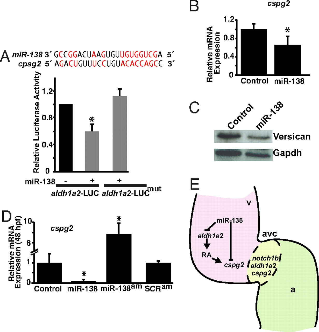
Fig. 5 miR-138 directly targets cspg2. (A) Sequence of miR-138 and its binding site in mouse cspg2 3′ UTR with complementary nucleotides indicated in red. Luciferase activity in Cos cells on introduction of wild type or mutated (mut) cspg2 3′ UTR sequences downstream of a CMV-driven luciferase (LUC) reporter with or without miR-138 is shown. (B and C) Analysis of cspg2 mRNA level assessed by qRT-PCR (B) and corresponding versican protein by western blot (C) in mouse NIH 3T3 fibroblasts transfected with miR-138. (D) cspg2 mRNA levels detected by qRT-PCR in zebrafish embryos injected with miR-138 or treated at 24 hpf with miR-138 antagomiR (am) or scrambled (SCR) antagomiR. (E) Proposed model for the function of miR-138 in regulating chamber-specific gene expression and atrioventricular canal (avc) patterning. miR-138 directly represses retinoic acid (RA) synthesis in the ventricle (v) via aldh1a2, which would otherwise induce expression of the avc-specific gene, cspg2 (versican). Repression of cspg2 by miR-138 in the ventricle is also accomplished by directly targeting cspg2. Results shown in (A), (B), and (D) represent at least 4 experiments with error bars indicating 95% confidence intervals. (a, atrium; *, P < 0.05.)