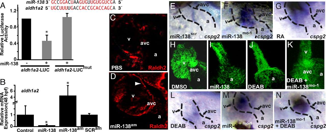
Fig. 4 miR-138 directly targets aldh1a2, restricting its expression to the AVC. (A) miR-138 binding site in zebrafish aldh1a2 3′ UTR with complementary nucleotides indicated in red. Luciferase activity in Cos cells on introduction of wild-type or mutated (mut) aldh1a2 3′ UTR sequences downstream of a CMV-driven luciferase reporter with or without miR-138 is shown. (B) aldh1a2 mRNA levels measured by qRT-PCR in embryos injected with pri-miR-138 or treated at 24 hpf with miR-138 antagomiR (miR-138am) or scrambled antagomiR (SCRam). Results shown represent at least 4 experiments in (A) and (B). Error bars indicate 95% confidence intervals. (C and D) Raldh2 immunohistochemistry on sections of 72-hpf embryos treated with PBS or miR-138am from 24–72 hpf. Ventricular (v) cardiomyocyte expression (arrowhead) was readily detectable in miR-138am hearts but not in controls, whereas intensity of staining in the AVC was similar. (E–G) Ventral view of cspg2 expression in the heart by mRNA in situ hybridization in embryos treated with scrambled morpholino (miR-138scr) (E), miR-138mo-1 (F), or retinoic acid (RA) (G); heart is indicated with dotted lines. (H–K) Confocal images of myl7-GFP embryos (48 hpf) treated with vehicle (DMSO) (H), premiR-138 RNA (I), or DEAB (J), and DEAB-treated embryos also injected with miR-138mo-1 (K) showing rescue of the linear heart tube phenotype with knockdown of miR-138. (L–N) Expansion of cspg2 mRNA expression from the avc (L) into the ventricle (v) of miR-138mo-1 embryos (M) was not rescued by DEAB (N). (a, atrium; *, P < 0.05.)
Image
Figure Caption
Acknowledgments
This image is the copyrighted work of the attributed author or publisher, and
ZFIN has permission only to display this image to its users.
Additional permissions should be obtained from the applicable author or publisher of the image.
Open Access.
Full text @ Proc. Natl. Acad. Sci. USA

