
|
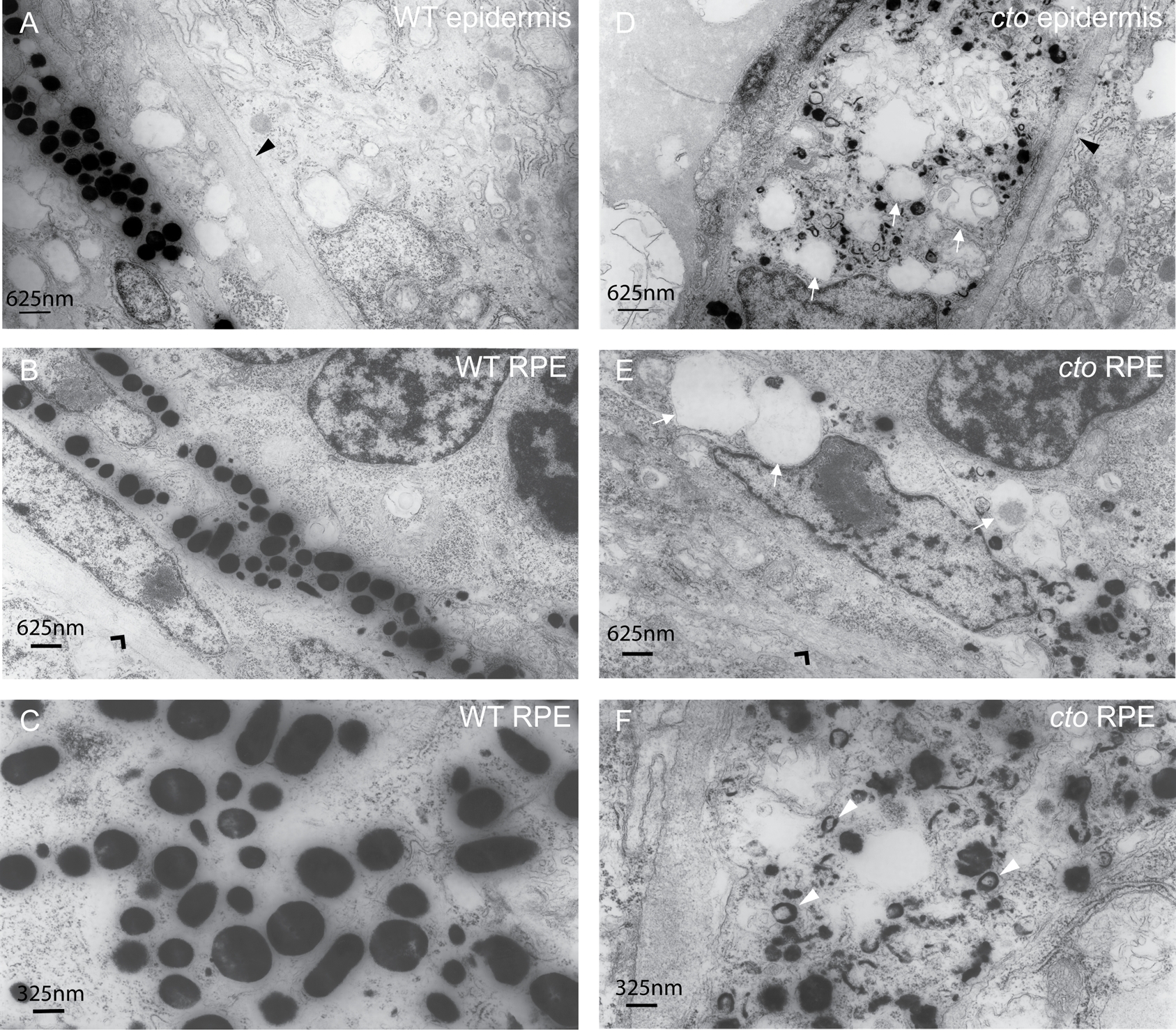
Fig. 6 cto melanocytes have significant ultrastructural defects.
(A–B) Wild-type epidermal (A) and retinal pigment epithelial (RPE, B) melanocytes are elongated and thin and contain many large, densely pigmented melanosomes. The epithelial basement membrane is indicated by a black triangle in (A) and the RPE basement membrane by a black arrowhead in (B). (C) A higher magnification of wild-type melanosomes showing significant pigmentation and ellipsoid shape when cut longitudinally. (D–E) cto mutant epidermal (D) and RPE (E) melanocytes showing rounded, poorly pigmented cells that contain numerous large, empty vesicles (white arrows) The basement membranes are indicated as in (A and B). (F) A higher magnification of RPE melanosomes showing the diverse array of immature, poorly pigmented vesicles. The white arrowheads point to multi-lamellar, melanin filled vesicles which are identical to the melanin positive multi-vesicular bodies seen in the cappucino mouse (see text).