
Fig. S2
- ID
- ZDB-IMAGE-081117-24
- Publication
- Chung et al., 2008 - Bmp2 signaling regulates the hepatic versus pancreatic fate decision
- All Figures
- Figures for Chung et al., 2008
|
Fig. S2
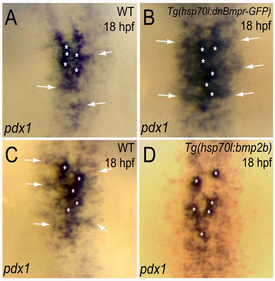
Bmp Signaling Regulates pdx1 Expression
(A–D) Dorsal views of pdx1 expression, comparing wild-type (A and C), Tg(hsp70l:dnBmpr-GFP)w30 (B) and Tg(hsp70l:bmp2b)f13 (D) hemizygous embryos at 18 hpf. pdx1 is expressed at high levels in the most medial cells (asterisks) and at low levels in the lateral cells (arrows) in wild-type (A and C). (B) In Tg(hsp70l:dnBmpr-GFP)w30 hemizygous embryos heat-shocked at the 6-somite stage, pdx1 expression was expanded laterally (the percentage of embryos showing a substantial lateral expansion of pdx1 expression was 60%, n = 18/31). (D) In Tg(hsp70l:bmp2b)f13 hemizygous embryos heatshocked at the 8-somite stage, high levels of pdx1 expression (asterisks) were maintained, while low levels (arrows in A, B and C) were severely reduced (the percentage of bmp2b).
Reprinted from Developmental Cell, 15(5), Chung, W.S., Shin, C.H., and Stainier, D.Y., Bmp2 signaling regulates the hepatic versus pancreatic fate decision, 738-748, Copyright (2008) with permission from Elsevier. Full text @ Dev. Cell