
Fig. 3
- ID
- ZDB-IMAGE-081111-8
- Publication
- Linney et al., 1999 - Transgene expression in zebrafish: a comparison of retroviral-vector and DNA-injection approaches
- All Figures
- Figures for Linney et al., 1999
|
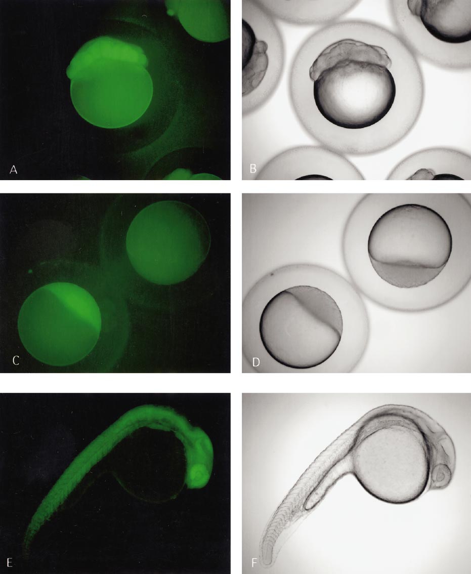
Fig. 3 GFP-positive embryos derived from transgenic founder produced via pseudotyped retroviral vector infection. (A and B) 16-cell embryos derived from transgenic female parent—note the GFP even though the zygotic genome has not turned on. (C and D) GFP from a dome-stage embryo—the transgenic embryo in the lower left was derived from a transgenic female parent while the transgenic embryo in the upper right was derived from a transgenic male parent. The fluorescence in the lower left embryo is derived from the oocyte. Even though the zygotic genome is beginning to turn on at this time, we routinely do not detect embryonic GFP until 5–7 h after fertilization. (E and F) 24-h embryo derived from transgenic female parent.
Reprinted from Developmental Biology, 213(1), Linney, E., Hardison, N.L., Lonze, B.E., Lyons, S., and DiNapoli, L., Transgene expression in zebrafish: a comparison of retroviral-vector and DNA-injection approaches, 207-216, Copyright (1999) with permission from Elsevier. Full text @ Dev. Biol.