
|
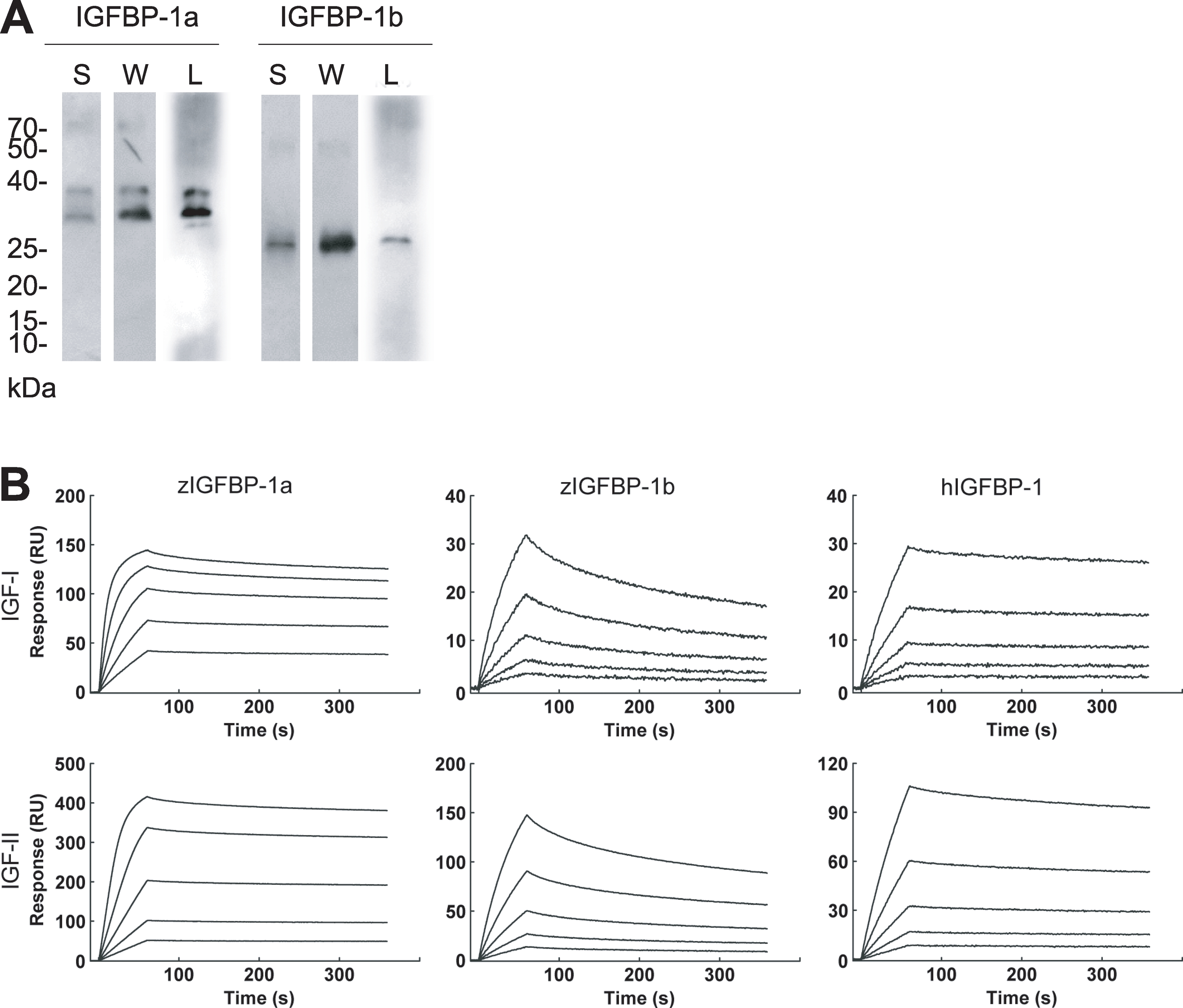
Fig. 5
Zebrafish IGFBP-1a and -1b proteins have different IGF binding affinities and kinetics.
A) Purification and characterization of zebrafish IGFBP-1a and -1b proteins. Isolated IGFBP-1a and -1b proteins were analyzed by SDS-PAGE under non-reducing condition, followed with Silver staining (S), western blotting (I) and western ligand-blotting (L). 1.0 (S) or 0.3 μg (W and L) purified protein was loaded to each lane. (B) BIAcore analysis of IGFBP1a and -1b. All binding experiments were carried out at 25°C in HBS buffer with a constant flow rate of 30 μL/min. Different concentrations of zebrafish IGFBP-1a and -1b or human IGFBP-1 were injected over the surface for 60 seconds followed by a five minute dissociation period. The sensor surface was regenerated by two 30 second injections of HCl (100 mM). Sensorgram data were analyzed using the BIAcore T100 evaluation Software version 1.1. Concentrations of IGFBP-1a applied were 144 nM, 72 nM, 36 nM, 18 nM and 9 nM, IGFBP-1b were 173 nM, 86.4 nM, 43.2 nM, 21.6 nM and 10.8 nM and human IGFBP-1 were 368 nM, 184 nM, 92 nM, 46 nM and 23 nM.