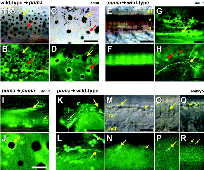
Fig. 11 Cell transplantation reveals an essential role for puma in promoting multiple cell lineages during postembryonic development. All images are whole mounts. Red arrows, wild-type cells. Yellow arrows, puma mutant cells. (A–D) Wild-type cells transplanted into puma hosts contributed to a broad array of derivatives (Table 1), including pigment cells, when examined at adult stages. (A, B) Corresponding brightfield and fluorescence views. Many chimeras developed donor-derived GFP+ wild-type pigment cells. Large red arrow, example of a donor-derived wild-type melanophore. Although donor melanophores could be spread widely over the flank, they were typically interspersed with host GFP- puma mutant melanophores, which retained their characteristic large size as compared with wild-type melanophores (yellow arrow). Donor wild-type cells also differentiated into placode-derived tissues and components of the peripheral nervous system: small red arrow, posterior trunk lateral line nerve. (C, D) Corresponding brightfield and fluorescence views. Higher magnification view of a different individual showing donor wild-type GFP+ melanophores (red arrow) adjacent to host puma mutant melanophores (yellow arrow). Note that melanin (contained within melanosomes) remains partially dispersed in the peripheries of the puma mutant melanophores despite treatment with epinephrine, whereas melanin in wild-type melanophores is contracted toward the cell centers. (E–H) puma mutant cells transplanted into wild-type hosts contributed to only a limited array of derivatives (Table 1) when examined at adult stages. (E, F) Corresponding brightfield and fluorescence views showing donor GFP+ puma mutant contribution to vertebrae (v). nt, neural tube; a, aorta. A few wild-type host melanophores are visible in the background above the neural tube and below the aorta. (G) Donor puma mutant cells that differentiated as muscle fibers within the myotome (arrow). (H) In a single chimera, donor puma mutant cells differentiated as a single identifiable neuron (yellow arrow). Fluorescence from adjacent host GFP- xanthophores (large red arrow) is color-shifted relative to GFP fluorescence. Note absence of GFP fluorescence in host melanophore as well (small red arrow). (I, J) puma mutant cells contribute to a broader spectrum of derivatives when transplanted to puma mutant hosts and examined in the adult. (I) Donor GFP+ puma mutant cells can be seen within the myotome (large arrow) and the vertebral column (small arrow). (J) puma mutant cells also contribute to pigment cell lineages in puma mutant hosts, including melanophores (arrow), in contrast to the behavior of these cells in wild-type hosts. (K–R) puma mutant cells are able to produce a wide variety of derivatives at embryonic stages, when transplanted to wild-type embryos. Shown are embryos at 25–27 hpf shortly after melanophores have started to acquire melanin. (K) Head of an embryo showing donor GFP+ puma mutant cells contributing to the eye (small arrow), epidermis, and posterior lateral line nerve (large arrow, enlarged in L). (L) puma mutant donor cells contributing to the migrating posterior lateral line nerve (large arrow) as well as epidermis (small arrow). (M–R) puma mutant cells also are present within the premigratory neural crest and in neural crest migratory pathways, though these cells are often irregularly shaped. (M, N) Corresponding brightfield and fluorescence views of a donor GFP+ puma mutant cell within the epidermis dorsal to the neural crest (large arrow); morphology resembles that of typical zebrafish apoptotic bodies (Parichy et al 1999 and Parichy and Turner 2003b). Small arrow, lightly melanized host wild-type melanophore. (O, P) GFP+ puma mutant cell within the dorsolateral neural crest migratory pathway is irregularly shaped and also resembles typical apoptotic body. (Q, R) Punctate GFP+ puma mutant cells within the neural crest. (A, B) 200 μm; (C, D) 100 μm; (E, F) 200 μm; (G) 100 μm; (H) 60 μm; (I, J) 100 μm; (K, L) 40 μm; (M, N) 40 μm; (O–R) 20 μm.
Reprinted from Developmental Biology, 256(2), Parichy, D.M., Turner, J.M., and Parker, N.B., Essential role for puma in development of postembryonic neural crest-derived cell lineages in zebrafish, 221-241, Copyright (2003) with permission from Elsevier. Full text @ Dev. Biol.

