
Fig. 6
- ID
- ZDB-IMAGE-081021-73
- Publication
- Lekven et al., 2003 - Wnt1 and wnt10b function redundantly at the zebrafish midbrain-hindbrain boundary
- All Figures
- Figures for Lekven et al., 2003
|
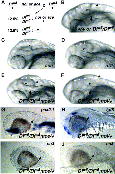
Fig. 6 ace and noi are dominant enhancers of Dfw5. (A) Schematic diagram of the cross performed to generate embryos homozygous for Dfw5 and heterozygous for ace or noi. (B–F) Oblique views of heads of live 27-hpf embryos. Genotypes are indicated in the lower right corners. (B) In wild type or Dfw5 homozygotes, the fold is visible and creates a clear separation between the posterior edge of the tectum (arrowhead) and the cerebellum (small arrow). The midline of the fold (large arrow) is visible as an indentation in the lateral hindbrain wall. Asterisks in all panels: dorsal midline of the midbrain. (C) ace mutants fail to form a MHB fold and do not form a cerebellum. Rather, the posterior edge of the optic tectum is in a position corresponding to the position of the cerebellum in wild types (arrowhead). (D) noi mutants also fail to form the MHB fold and cerebellum, but also have defects in the formation of the midbrain. Thus, the tectum appears much smaller than in ace mutants and also has a turbid appearance (not visible). Dfw5 mutants enhanced by ace (E) or noi (F) alleles appear similar in that the separation between the tectum and cerebellum is less distinct due to the absence of a definitive MHB fold. Arrowheads indicate the posterior edge of the tectum, and small arrows indicate the position of the cerebellum. A very slight bump in the hindbrain wall is visible (large arrow). (G–J) Loss of MHB gene expression in Dfw5 embryos enhanced by ace (G, I) or noi (H, J). Probe used is indicated in the upper right corner. Note the absence of expression of each gene in the MHB region (arrows) in comparison to Dfw5 homozygotes (Fig. 4).
Reprinted from Developmental Biology, 254(2), Lekven, A.C., Buckles, G.R., Kostakis, N., and Moon, R.T., Wnt1 and wnt10b function redundantly at the zebrafish midbrain-hindbrain boundary, 172-187, Copyright (2003) with permission from Elsevier. Full text @ Dev. Biol.