
Fig. 9
- ID
- ZDB-IMAGE-081021-56
- Publication
- Huang et al., 2003 - reg6 is required for branching morphogenesis during blood vessel regeneration in zebrafish caudal fins
- All Figures
- Figures for Huang et al., 2003
|
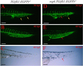
Fig. 9 Branching defects in developing caudal veins in reg6 embryos. (A) A wild-type TG(fli1:EGFP)y1 embryo at 30 h postfertilization (hpf) (33°C) shows numerous branches (yellow arrows) in the developing caudal vein. (B) These branches develop into a plexus within a few hours and the plexus persists for several days during embryogenesis (a 40-hpf embryo is shown). (C) Bright field image of (B). (D) The developing caudal vein in reg6;TG(fli1:EGFP)y1 embryo at 30 hpf (33°C) shows fewer branches (yellow arrow) and a developing sinus (red arrow). (E) By 40 hpf, the developing caudal veins in reg6;TG(fli1:EGFP)y1 embryos become swollen and form a sinus (red arrow). (F) Bright field image of (E); red arrow, developing sinus. Anterior to the left and dorsal up. hpf, hour post fertilization. Scale bars, 100 μm.
Reprinted from Developmental Biology, 264(1), Huang, C., Lawson, N.D., Weinstein, B.M., and Johnson, S.L., reg6 is required for branching morphogenesis during blood vessel regeneration in zebrafish caudal fins, 263-274, Copyright (2003) with permission from Elsevier. Full text @ Dev. Biol.