
Image
|
Figure Caption
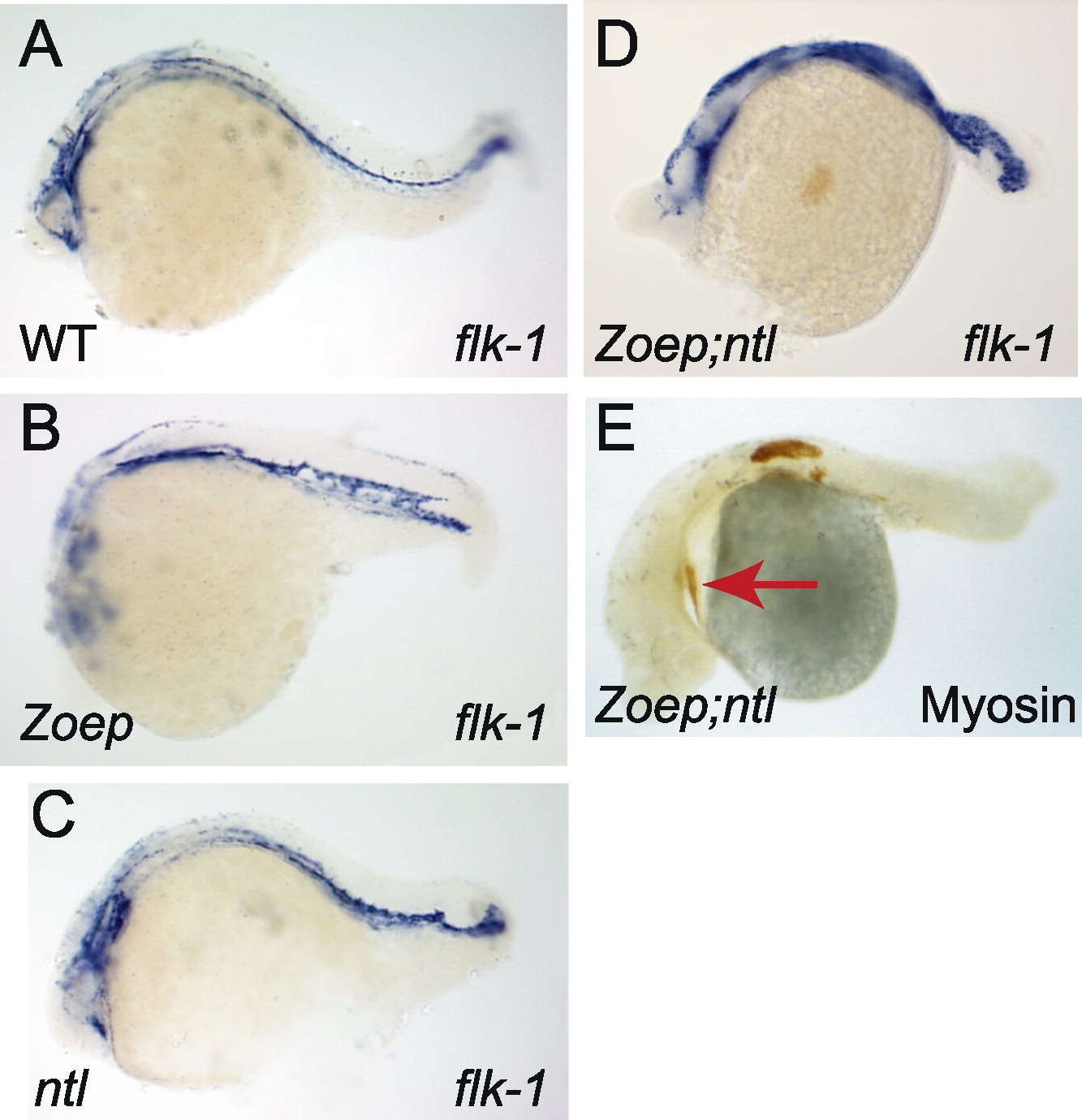
Fig. 1 Zoep and ntl are not required for the formation of cardiac mesoderm or vascular endothelium. Anterior, left; dorsal, uppermost; genotypes as indicated (bottom left) (A–D) flk-1 expression. Note the presence of flk-1 expressing cells in the Zoep;ntl embryo, which are disorganized and possibly more numerous than in wild-type or either single mutant embryos. (E) Myosin staining (brown) detects the cardiac primordium (arrow), as well as somitic tissue in the anterior trunk, as previously reported.
Acknowledgments
This image is the copyrighted work of the attributed author or publisher, and
ZFIN has permission only to display this image to its users.
Additional permissions should be obtained from the applicable author or publisher of the image.
Reprinted from Developmental Biology, 264(2), Griffin, K.J. and Kimelman, D., Interplay between FGF, one-eyed pinhead, and T-box transcription factors during zebrafish posterior development, 456-466, Copyright (2003) with permission from Elsevier. Full text @ Dev. Biol.