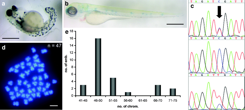
Fig. 3 Progeny of mlh1 -/- males. a Severely malformed 3-day-old embryo from a cross of a mlh1 -/- male with a wild-type female. b Unpigmented 3-day-old embryo from a cross of a mlh1 -/- male with an albino female, showing that the wild-type paternal allele of the albino gene has been lost in this embryo. The weak blue staining is caused by the methylene-blue-containing medium. c Sequence traces of the polymorphism in mlh1 (arrow). The paternal allele (mutant) is a T, and the maternal allele (wild-type) is a C. The sequence peak areas show quantitatively that some progeny of mutant males have one chromosomal copy of each parent (top), whereas others have only the maternal allele (middle) or two copies of the paternal allele (bottom that some progeny of mutant males have one chromosomal copy of each parent (top), whereas others have only the ma). d Embryos have chromosome numbers different from the normal number of 50, such as this example of 47 chromosomes. e Distribution of chromosome numbers in embryos (emb.) from mlh1 -/- males (30 embryos in total). Bars 500 μm (a, b), 1 μm (d)
Image
Figure Caption
Figure Data
Acknowledgments
This image is the copyrighted work of the attributed author or publisher, and
ZFIN has permission only to display this image to its users.
Additional permissions should be obtained from the applicable author or publisher of the image.
open access
Full text @ Cell Tissue Res.

