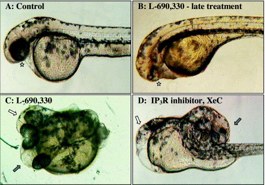
Fig. 5 Window of sensitivity and axis duplication induced by PI cycle inhibition. Lateral view with anterior to the left of live embryos after 48 hpf. Late treatment of zebrafish embryos with PI cycle inhibitors results in anterior head defects and fusion of the eyes (U-73122, 2.4%; XeC, 3.2%; and L-690,330, 5.7%). (A) Control embryo with star denoting one eye in focus. (B) Embryo injected at a late stage with L-690,330 with the star denoting a reduced eye with partial cyclopia. PI cycle inhibition generates duplicated dorsal structures. (C) L690,330-injected embryo with duplicated heads (arrows). The fourth eye is slightly out of the focal plane (white arrow). (D) XeC-injected embryo with the partial secondary axis including ectopic ears (white arrow) and abnormal pericardial cavity. The primary axis folded over the yolk (black arrow).
Reprinted from Developmental Biology, 259(2), Westfall, T.A., Hjertos, B., and Slusarski, D.C., Requirement for intracellular calcium modulation in zebrafish dorsal-ventral patterning, 380-391, Copyright (2003) with permission from Elsevier. Full text @ Dev. Biol.

