
|
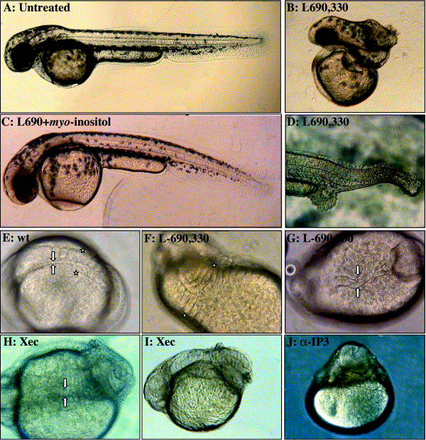
Fig. 2 Range of morphological phenotypes induced by PI cycle inhibition. Lateral view with anterior to the right of live embryos approximately 48 hpf (A–D). (A) Untreated. (B) L-690,330-injected (3–6 mM) with reduced tail tissue (similar to lost-a-fin, C2). (C) control L-690,330 (10 mM) + myo-inositol-injected (50 mM). (D) L-690,330-injected (10 mM, snail-house-like, C4). Dorsal (E–G) and lateral (I–J) view of ∼24 hpf embryos. (E) Wild-type embryo with arrows denoting notochord width and stars highlight the lateral portion of one pair of somites. L-690,330-injected embryos display both (F) expanded somites, stars and (G) thickened notochord, arrows similar to swirl-like phenotypes (C5). Embryos treated with IP3Receptor inhibitors display (H) thickened, wavy notochord (arrows, XeC). (I) tail curling phenotypes similar to piggy-tail-like (C3) (XeC), and (J) monoclonal blocking antibody-induced bustling and radialization of tissue over the yolk.
Reprinted from Developmental Biology, 259(2), Westfall, T.A., Hjertos, B., and Slusarski, D.C., Requirement for intracellular calcium modulation in zebrafish dorsal-ventral patterning, 380-391, Copyright (2003) with permission from Elsevier. Full text @ Dev. Biol.