Fig. 8
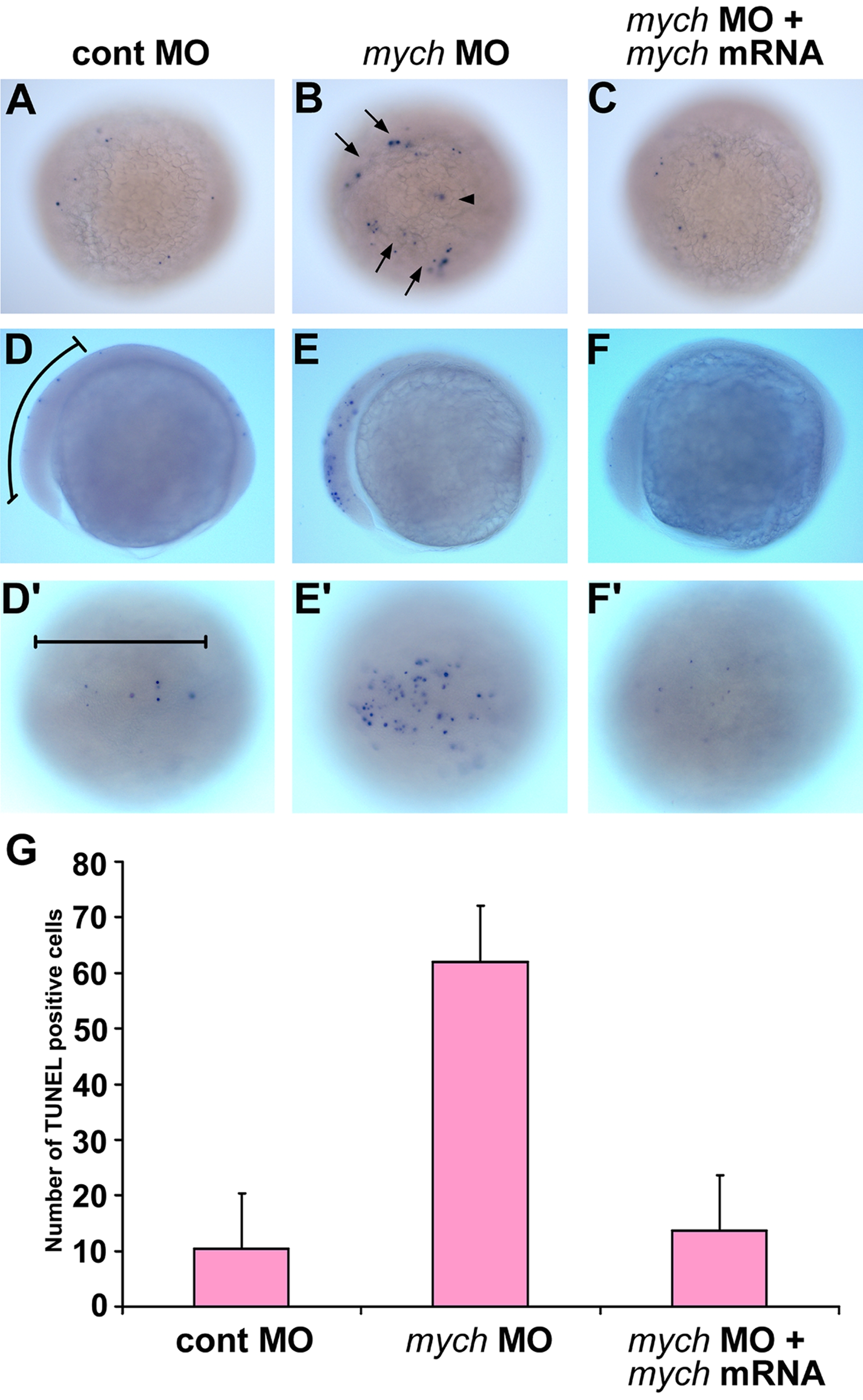
Mych depletion results in apoptosis in the early neural plate.
A–F′. Detection of cell death by TUNEL assay. Lateral views (D,E,F) and dorsal views (A,B,C,D′,E′,F′) of control MO (A,D,D′), mych UTR MO (B,E,E′), and rescued embryos that received mych UTR MO and mRNA (C,F,F′) at the bud (A–C) and 3-somite stage (D–F′). Arrows in B point out TUNEL positive cells at the lateral edge of the neural plate. An arrowhead in B indicates TUNEL positive cells in the neural plate. Bracket in D and D′ indicates the anterior neural plate region in which TUNEL-positive cells were counted. The results are shown in G. Average numbers of positive cells per embryo were obtained by counting 20 embryos in each group. For both comparisons p<0.01: Cont MO vs. mych MO: p = 5.27e-14; mych MO vs. rescue: p = 4.87e-17.
Image
Figure Caption
Figure Data
Acknowledgments
This image is the copyrighted work of the attributed author or publisher, and
ZFIN has permission only to display this image to its users.
Additional permissions should be obtained from the applicable author or publisher of the image.
Open Access
Full text @ PLoS One

