
Fig. S4
- ID
- ZDB-IMAGE-081007-28
- Genes
- Publication
- Mudumana et al., 2008 - odd skipped related1 reveals a novel role for endoderm in regulating kidney versus vascular cell fate
- All Figures
- Figures for Mudumana et al., 2008
|
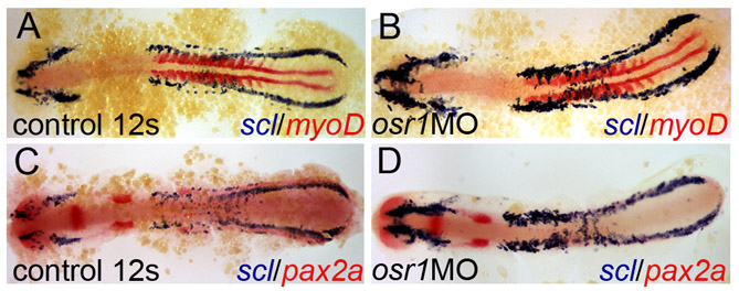
Fig. S4 Expanded vascular differentiation is not due to an altered rate of development. (A) scl (blue) and myoD (red) expression in wild-type embryos at the 12-somite stage. (B) osr1 knockdown results in expansion of scl-positive tissue, most prominently in anterior PLM. myoD is used as an internal staging control and demonstrates both control and osr1 morphants are at the 12 somite stage. (C) scl (blue) and pax2a (red) expression in wild-type embryos from the same clutch of embryos as A and B. (D) osr1 knockdown results in expansion of scl-positive tissue, most prominently in anterior PLM, and loss of pax2a-expressing cells. (C, D) are lower magnification views of A,B in Fig. 5 respectively.