
Fig. 1
- ID
- ZDB-IMAGE-080925-2
- Genes
- Antibodies
- Publication
- Del Bene et al., 2008 - Regulation of neurogenesis by interkinetic nuclear migration through an apical-basal notch gradient
- All Figures
- Figures for Del Bene et al., 2008
|
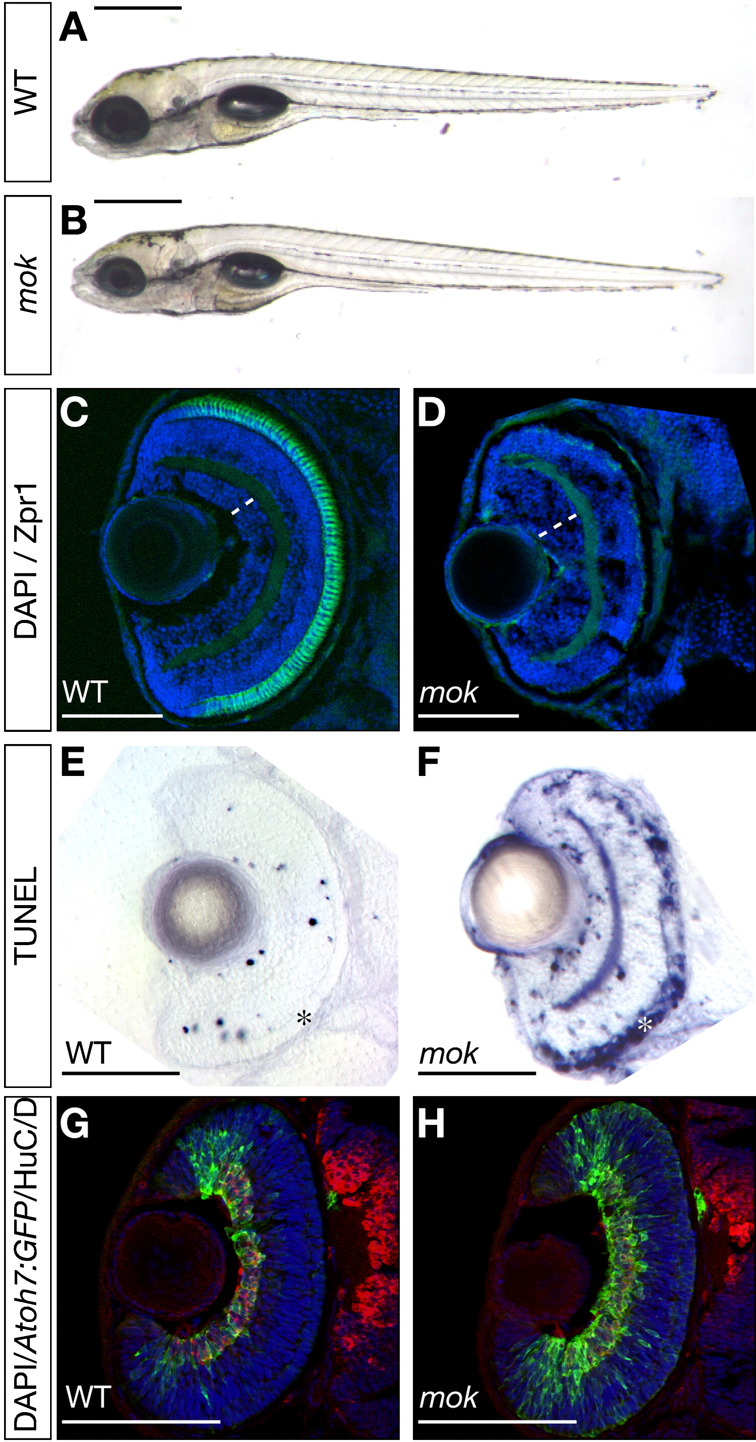
Fig. 1 moks309 Mutants Have a Complex Retina Phenotype
(A and B) Whole-mount lateral views of live 4.5 dpf zebrafish larvae. In moks309 mutants (B), the eyes are smaller than in the wild-type (A), but external morphology is otherwise indistinguishable.
(C and D) Horizontal sections of 5 dpf retinas stained with DAPI (blue) and Zpr1 antibody (green). moks309 retinas (D) have an expanded GCL (dashed white lines), compared to wild-type (C), and no photoreceptors, as revealed by Zpr1 staining.
(E and F) Coronal sections of 3 dpf retinas stained by TUNEL assay. moks309 retinas (F) have increased apoptosis, particularly in the photoreceptor layer (asterisks), compared to wild-type (E).
(G and H) Horizontal sections of 48 hpf retinas expressing GFP driven by the atoh7 promoter (green) and stained with DAPI (blue) and HuC/D antibody (red). moks309 retinas (H) have an increased number of GFP-expressing RGCs compared to wild-type (G).
Scale bars, 500 μm (A and B) and 100 μm (C–H).
Reprinted from Cell, 134(6), Del Bene, F., Wehman, A.M., Link, B.A., and Baier, H., Regulation of neurogenesis by interkinetic nuclear migration through an apical-basal notch gradient, 1055-1065, Copyright (2008) with permission from Elsevier. Full text @ Cell