
Fig. S8
- ID
- ZDB-IMAGE-080925-16
- Publication
- Del Bene et al., 2008 - Regulation of neurogenesis by interkinetic nuclear migration through an apical-basal notch gradient
- All Figures
- Figures for Del Bene et al., 2008
|
Fig. S8
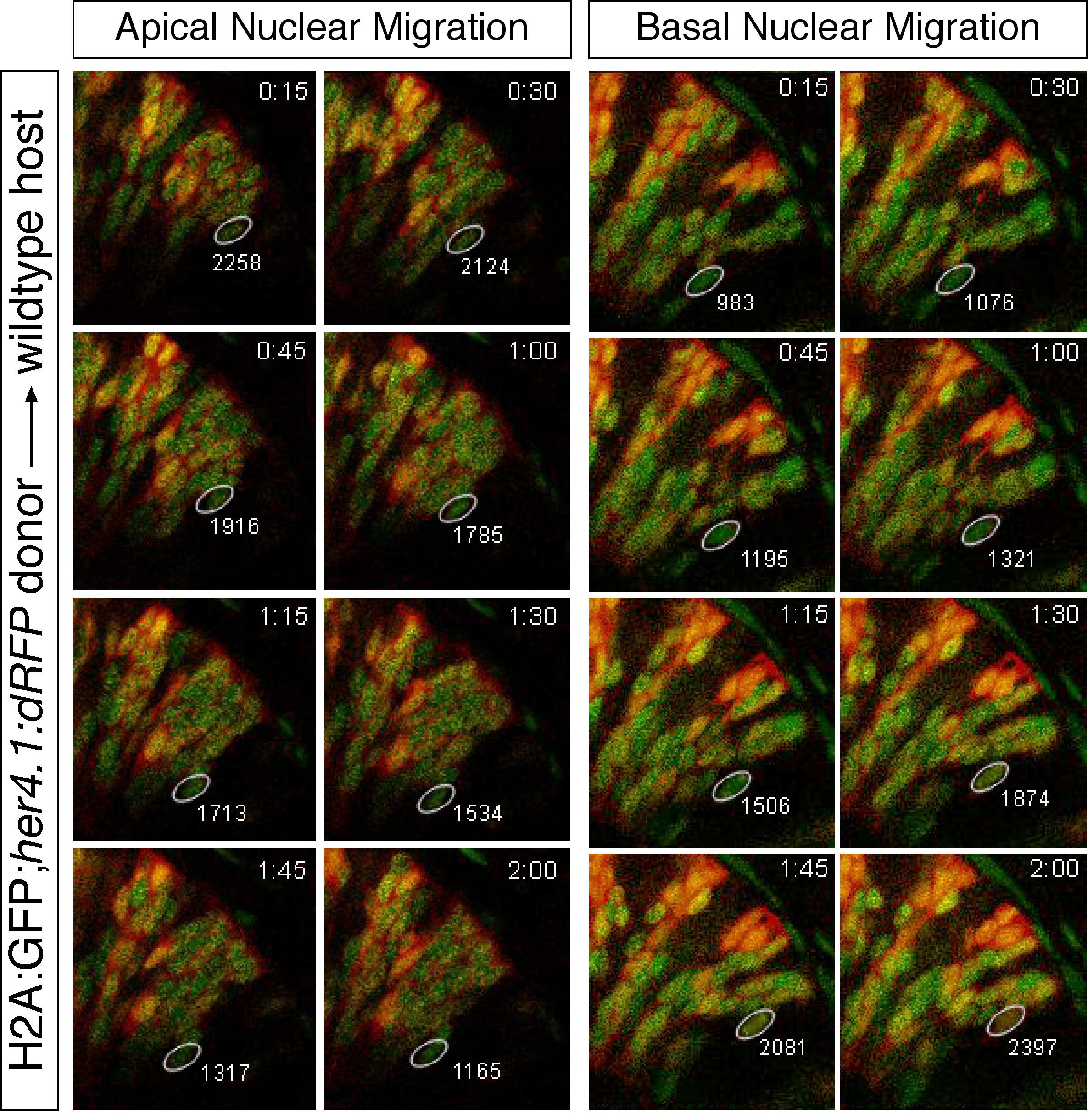
Retinal Neuroepithelia Show Changes in Notch Activity with Nuclear Migration
A. Montage of selected confocal planes showing decrease of the fluorescence signal in her4:dRFP pixel intensity with basal-directed nuclear migration.
B. Montage of the same cell later in development showing increased pixel intensity with apical-directed migration. In 11/15 cells moving nuclei apical to basal dRFP intensity decreased, while in 13/15 cells moving nuclei from basal to apical dRFP intensity increased. Time (hr:min) from initial pixel measurements is indicated in upper right. Integrated pixel intensity from the 543nm channel is indicated next to the region of interest. Apical surface is located in the upper right of each panel, while basal surface is to the lower left.
Reprinted from Cell, 134(6), Del Bene, F., Wehman, A.M., Link, B.A., and Baier, H., Regulation of neurogenesis by interkinetic nuclear migration through an apical-basal notch gradient, 1055-1065, Copyright (2008) with permission from Elsevier. Full text @ Cell