
Fig. S7
- ID
- ZDB-IMAGE-080925-15
- Publication
- Del Bene et al., 2008 - Regulation of neurogenesis by interkinetic nuclear migration through an apical-basal notch gradient
- All Figures
- Figures for Del Bene et al., 2008
|
Fig. S7
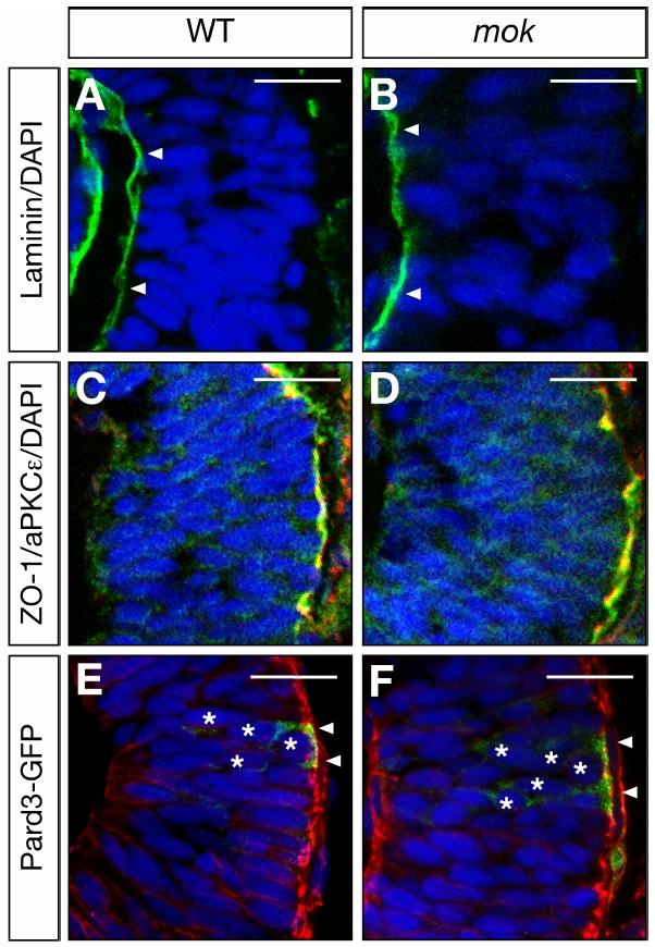
moks309 Mutants Have Normal Apical-Basal Marker Distribution
(A-D) 28 hpf retina sections stained with an antibody recognizing the basal lamina marker Laminin (green in [A, B]) and the apically distributed markers aPKCegreen in [C, D]) and the cell junction component ZO-1 (red in [C, D]). No obvious difference is detected between mutant (B, D) and wildtype (A, C) retinas.
(E,F) 28 hpf retina section stained with phalloidin (red) to detect a-actin and expressing Pard3-GFP (green). The majority of the Pard3-GFP protein is localized at the apical surface where it overlaps with the actin rich adherens-junctions. Nuclei of cells expressing the Pard3-GFP construct are marked by white stars. Nuclei counterstained with DAPI in blue. Scale bars 25 μm.
Reprinted from Cell, 134(6), Del Bene, F., Wehman, A.M., Link, B.A., and Baier, H., Regulation of neurogenesis by interkinetic nuclear migration through an apical-basal notch gradient, 1055-1065, Copyright (2008) with permission from Elsevier. Full text @ Cell