
Fig. 5
- ID
- ZDB-IMAGE-080923-5
- Antibodies
- Publication
- Tallafuss et al., 2008 - The Met receptor tyrosine kinase prevents zebrafish primary motoneurons from expressing an incorrect neurotransmitter
- All Figures
- Figures for Tallafuss et al., 2008
|
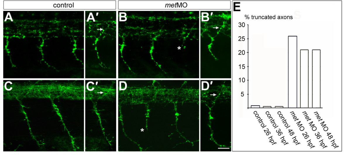
Fig. 5 Met is required for normal CaP axons. (a-b′) Embryos at 26 hpf showing PMN axons labeled with znp1 antibody. met MO-injected embryos have normal MiP axons [arrow in (b′)] compared to controls [arrow in (a′)]. However, some CaP axons are truncated in met MO-injected embryos [asterisk in (b)] compared to controls (a). (c-d′) Embryos at 48 hpf showing PMN and SMN axons labeled with znp1 antibody. As at 26 hpf, dorsally projecting axons in met MO-injected embryos appear normal [arrow in (d′)] compared to controls [arrow in (c′)]. However, some ventrally projecting axons are truncated in met MO-injected embryos [asterisk in (d)], compared to controls (c). (e) Average percentage of truncated axons in control and met MO-injected embryos at 26, 36 and 48 hpf. n (26 hpf) = 8 somites in each of 12 embryos; n (36 hpf) = 8 somites in each of 10 embryos; n (48 hpf) = 8 somites in each of 11 embryos. Scale bar, 20 μm.