
Fig. 7
- ID
- ZDB-IMAGE-080919-16
- Publication
- Fürthauer et al., 1999 - Three different noggin genes antagonize the activity of bone morphogenetic proteins in the zebrafish embryo
- All Figures
- Figures for Fürthauer et al., 1999
|
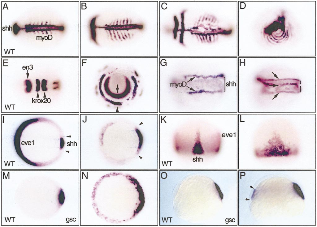
Fig. 7 Analysis of noggin1-injected embryos with molecular markers. All injected embryos received 20 pg nog1 mRNA. (A–H) 5-somite-stage embryos, dorsal views except (D) posterior view and (F) frontal view. (A) Uninjected wild-type control embryo. (B–D) In situ hybridization with shh, MyoD, and krox20 shows that following nog1 misexpression paraxial MyoD expression is enlarged and even circularized. (E, F) Expression of en3 at the midbrain–hinbrain boundary (arrow) and krox20 in rhombomeres 3 and 5 (arrowheads) ventrally encircle the embryo following nog1 overexpression. (G) Overexpression of nog1 can induce an enlargement of the axial mesoderm as revealed by expansion of the sonic hedgehog expression domain (bracket), lined by adaxial MyoD-expressing cells (arrows). (H) In 7% of the embryos the shh domain is split into two stretches separated by MyoD-expressing adaxial cells. (I–L) Embryos at midgastrula stages stained with eve1, a marker of the ventral margin, and shh, expressed in the axial mesendoderm. (I, J) Vegetal view, dorsal to the right. Following nog1 overexpression the eve1 expression domain is very much reduced, whereas the shh territory appears expanded. (K, L) Dorsal view. Nog1 overexpression enlarges the shh expression domain. (M–P) Shield-stage embryos, dorsal to the right. (M, N) Animal pole view. Nog1 overexpression induces gsc expression all around the margin of the blastoderm. (O, P) Lateral view. Ectopic gsc expression is limited to the marginal hypoblast (arrowheads).
Reprinted from Developmental Biology, 214(1), Fürthauer, M., Thisse, B., and Thisse, C., Three different noggin genes antagonize the activity of bone morphogenetic proteins in the zebrafish embryo, 181-196, Copyright (1999) with permission from Elsevier. Full text @ Dev. Biol.