
|
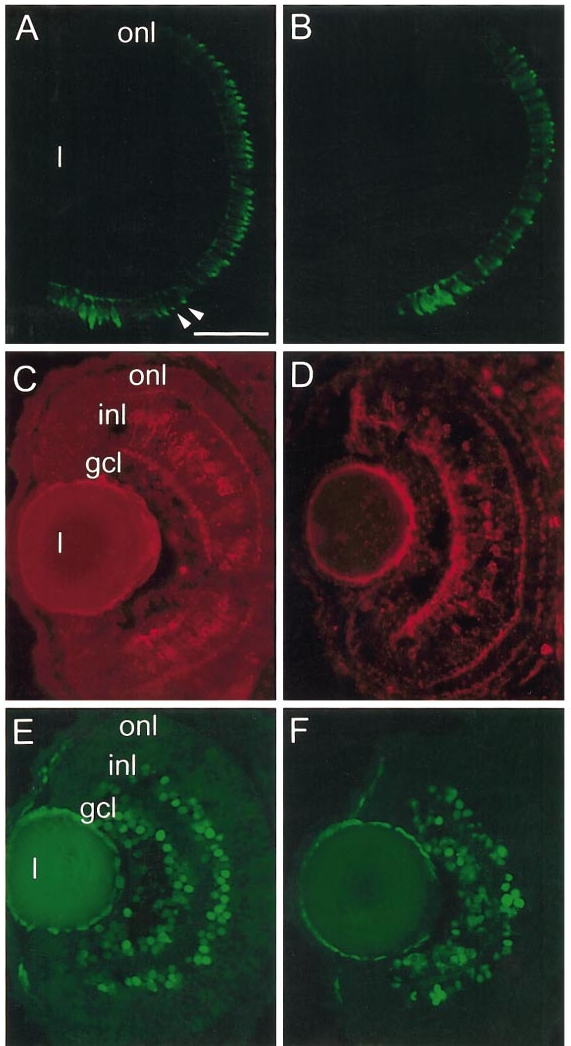
Fig. 6 Effect of shh/twhh antisense oligos on expression patterns of other retinal cell-specific markers. (A, C, and E) Sections obtained from embryos injected with the nonsense oligo. (B, D, and F) Sections obtained from embryos injected with the antisense oligo mixture. (A and B) Indirect immunofluorescence with zpr-1 (arrowheads indicate developing cone inner segments). (C and D) Indirect immunofluorescence using the anti-PKC antibody that labels rod bipolar cells. (E and F) Indirect immunofluorescence using the RET1 antibody that labels ganglion cells and a subpopulation of inner nuclear layer cells. l, lens; onl, outer nuclear layer; inl, inner nuclear layer; gcl, ganglion cell layer. Scale bar, 50 μm.
Reprinted from Developmental Biology, 220(2), Stenkamp, D.L., Frey, R.A., Prabhudesai, S.N., and Raymond, P.A., Function for hedgehog genes in zebrafish retinal development, 238-252, Copyright (2000) with permission from Elsevier. Full text @ Dev. Biol.