Image
Figure Caption
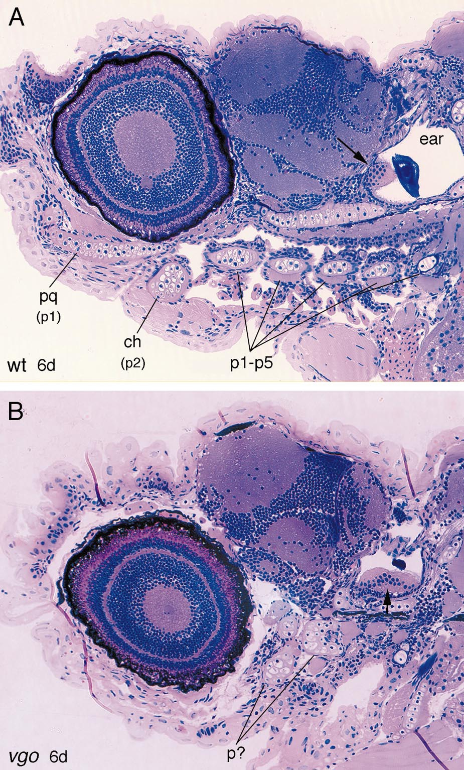
Fig. 6 2-μm-thick sagittal sections through 6-dpf wild-type (A) and vgo mutant larvae (B). In wild-type larvae the five posterior pharyngeal arches (p1–p5) are separated from one another by the gill clefts, whereas in vgo no organization of the arches is recognizable. Also the ear in vgo is much smaller, although the sensory patches (maculae, arrows) underlying the otoliths are present. Abbreviations: pq, palatoquadrate (dorsal element of first pharyngeal arch); p1, p2, pharyngeal arches 1 and 2; ch, ceratohyal (element of second pharyngeal arch).
Figure Data
Acknowledgments
This image is the copyrighted work of the attributed author or publisher, and
ZFIN has permission only to display this image to its users.
Additional permissions should be obtained from the applicable author or publisher of the image.
Reprinted from Developmental Biology, 225(2), Piotrowski, T. and Nüsslein-Volhard, C., The endoderm plays an important role in patterning the segmented pharyngeal region in zebrafish (Danio rerio), 339-356, Copyright (2000) with permission from Elsevier. Full text @ Dev. Biol.

