
|
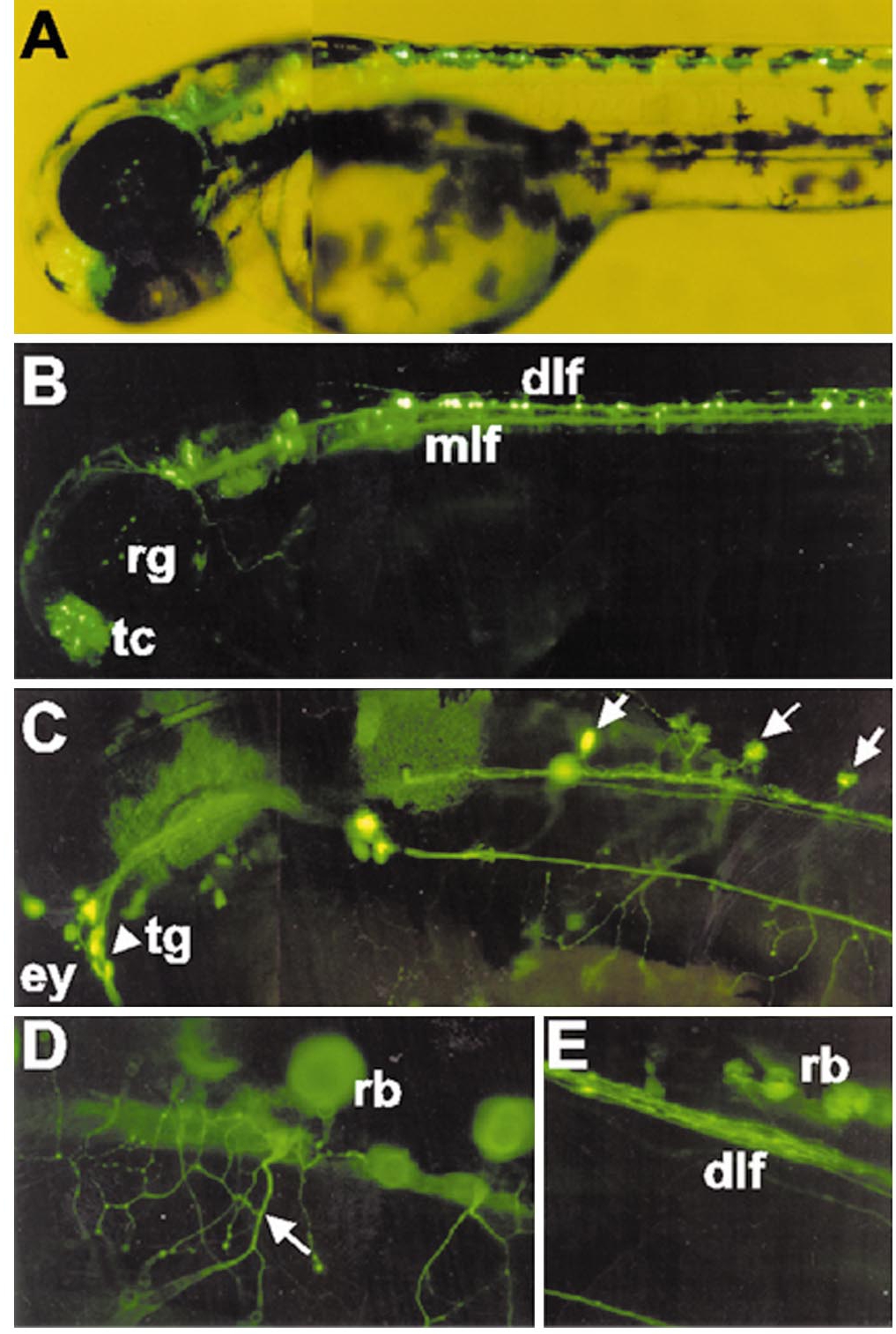
Fig. 5 Neuronal specificity of HuC promoter (ΔEco) in living zebrafish embryo. Neuronal specificity of transiently expressed GFP driven by the HuC promoter construct (ΔEco) in 48 hpf zebrafish embryos. (A) GFP fluorescence generated by superimposing a bright-field image on a fluorescence image. (B) Fluorescence image of GFP detected in the nervous system including the telencephalic cluster, retinal ganglion cells, medial longitudinal fasciculus, and dorsal longitudinal fasciculus. (C) GFP was expressed in the trigeminal ganglion neuron and Rohon-Beard neurons (arrow). (D, E) GFP expression in the peripheral process of Rohon-Beard axons (arrow) and dorsal longitudinal fasciculus of spinal cord. dlf, dorsal longitudinal fasciculus; ey, eye; mlf, medial longitudinal fasciculus; rb, Rohon-Beard neurons; rg, retinal ganglion; tc, telencephalic cluster; tg, trigeminal ganglion. Dorsal to the top and anterior to the left.
Reprinted from Developmental Biology, 227(2), Park, H.-C., Kim, C.-H., Bae, Y.-K., Yee, S.-Y., Kim, S.-H., Hong, S.-K., Shin, J., Yoo, K.-W., Hibi, M., Hirano, T., Miki, N., Chitnis, A.B., and Huh, T.-L., Analysis of upstream elements in the HuC promoter leads to the establishment of transgenic zebrafish with fluorescent neurons, 279-293, Copyright (2000) with permission from Elsevier. Full text @ Dev. Biol.