
IMAGE
Fig. S3
- ID
- ZDB-IMAGE-080910-19
- Antibodies
- Publication
- García-Lecea et al., 2008 - In vivo Analysis of Choroid Plexus Morphogenesis in Zebrafish
- All Figures
- Figures for García-Lecea et al., 2008
Image
|
Figure Caption
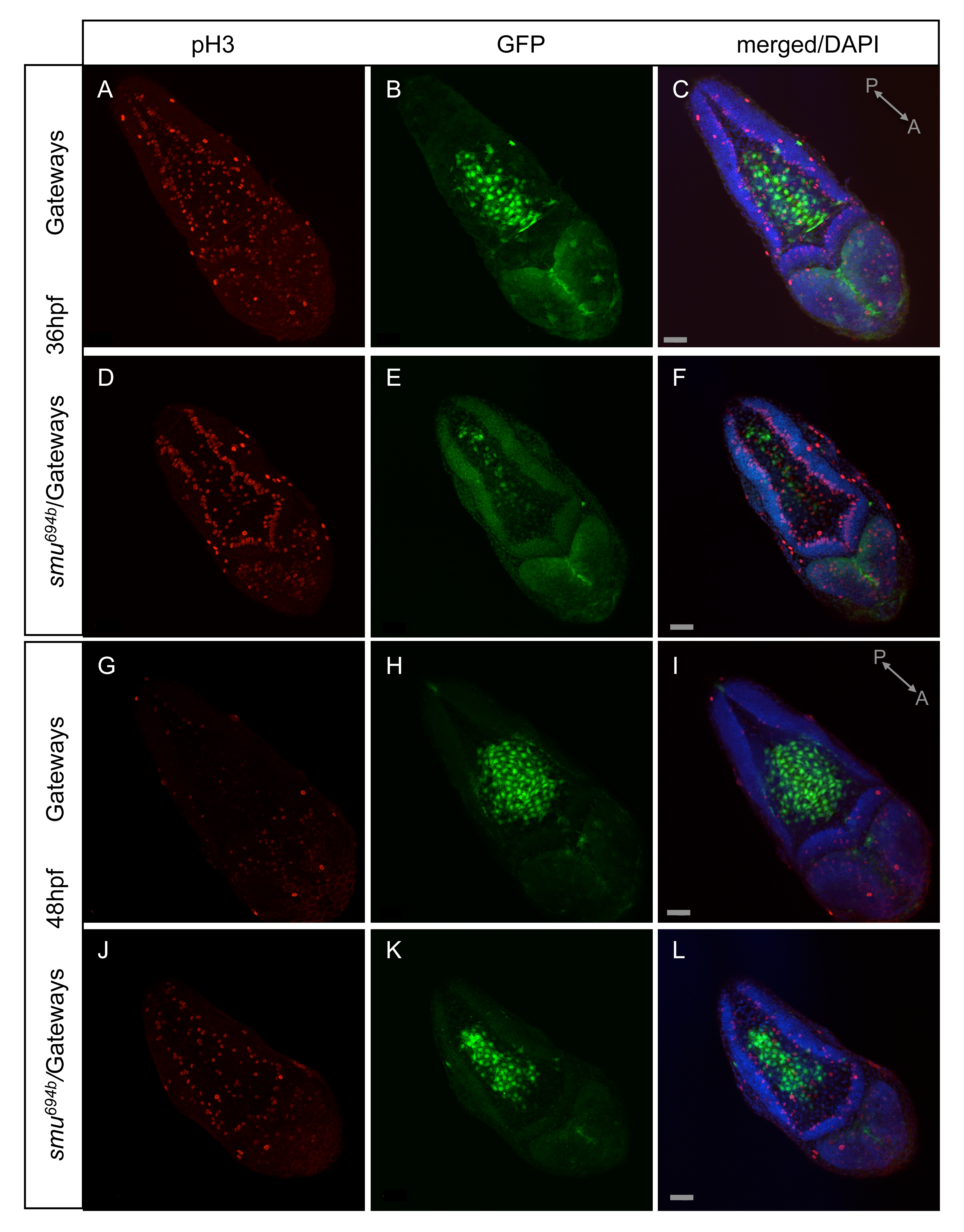
Fig. S3 Cell proliferation in the dorsal hindbrain as detected by anti-pH3 antibody. Two developmental stages were analyzed: 36hpf (A–C Gateways, D–F smu694b/Gateways) and 48hfp (G–I Gateways, J–L smu694b/Gateways). A,D,G,J - anti-pH3 antibody staining; B,E,H,K - anti-GFP antibody staining; C,F,I,L - merged images of anti-pH3/GFP staining and DAPI staining. All images are in dorsal view with anterior towards the right bottom corner.
Figure Data
Acknowledgments
This image is the copyrighted work of the attributed author or publisher, and
ZFIN has permission only to display this image to its users.
Additional permissions should be obtained from the applicable author or publisher of the image.
Full text @ PLoS One