
|
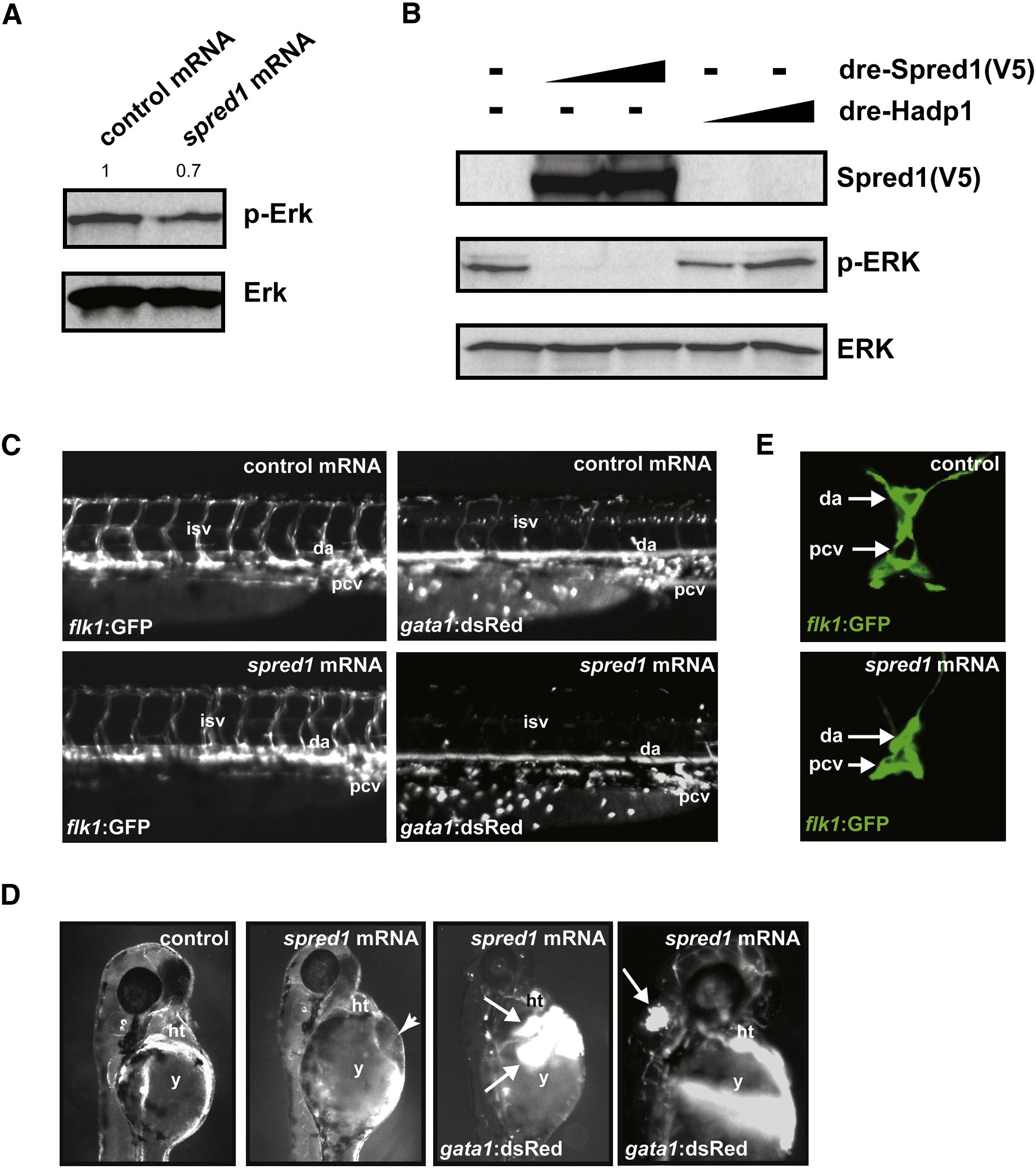
Fig. 6 Increased Spred1 Causes Vascular Instability and Hemorrhage Similar to miR-126 Knockdown
(A) Levels of phosphorylated Erk were reduced in zebrafish embryos (8 hpf) injected with spred1 mRNA compared to control mRNA (hadp1). Densitometric analysis is shown above.
(B) Zebrafish Spred1, but not Hadp1, expression also reduced p-ERK in COS cells as observed by immunoblot.
(C) Lateral view of trunk region of 48 hpf embryos after injecting 100 pg of control (hadp1) or spred1 mRNA. flk1:GFP reveals normal vascular patterning but gata1:dsRed shows diminished blood cells in the dorsal aorta (da), primary cardinal vein (pcv), and intersomitic vessels (isv).
(D) Injection of spred1 mRNA also resulted in pericardial (2nd panel from right) and cranial (far right) hemorrhages (arrows) visualized by gata1:dsRed marking of blood cells, and edema (2nd panel from left).
(E) Confocal analysis of transverse sections of 48 hpf embryos revealed collapsed lumens of da and primary cardinal vein (pcv) in spred1-injected embryos.
Reprinted from Developmental Cell, 15(2), Fish, J.E., Santoro, M.M., Morton, S.U., Yu, S., Yeh, R.F., Wythe, J.D., Ivey, K.N., Bruneau, B.G., Stainier, D.Y., and Srivastava, D., miR-126 regulates angiogenic signaling and vascular integrity, 272-284, Copyright (2008) with permission from Elsevier. Full text @ Dev. Cell