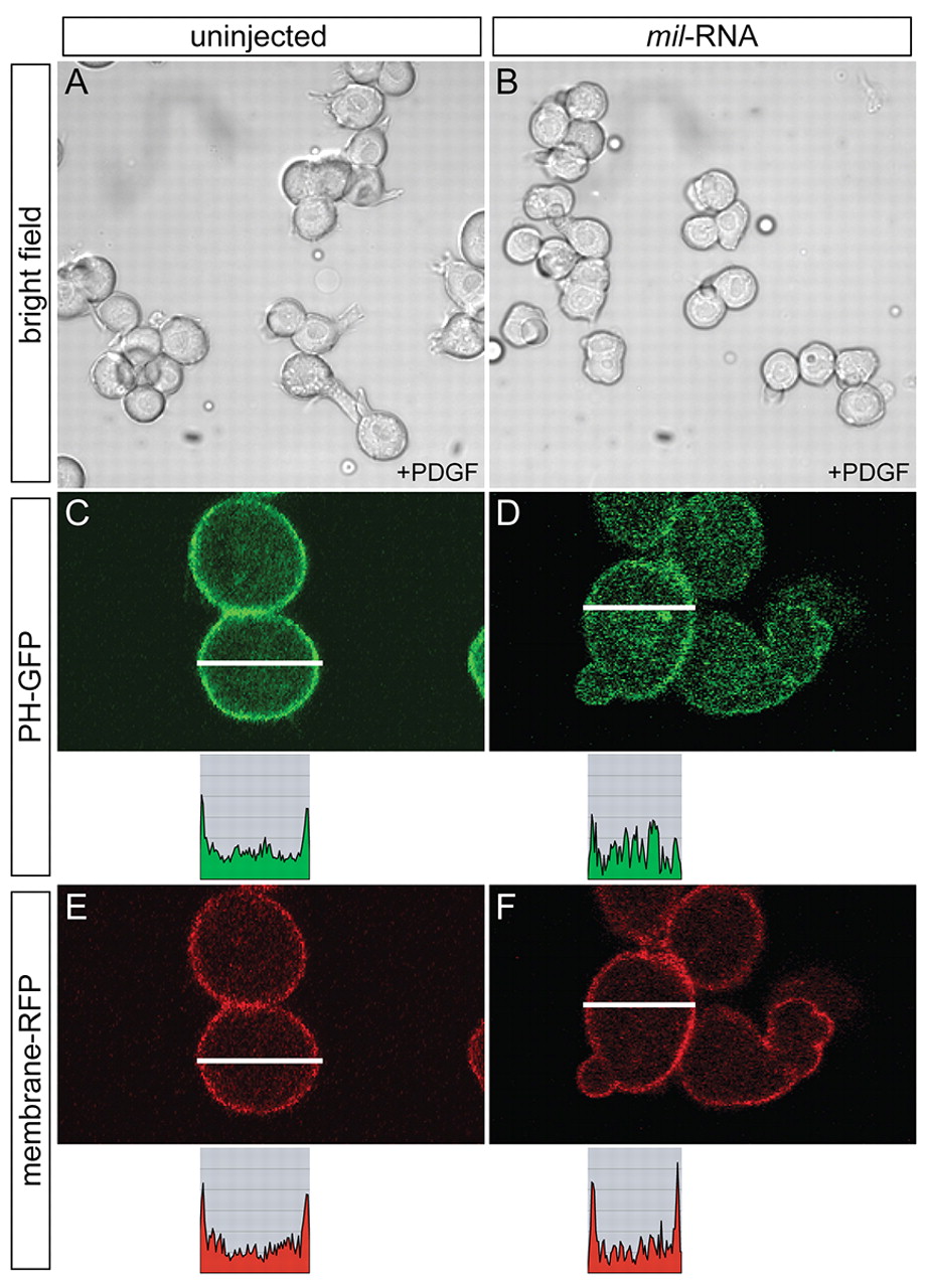
Fig. 7 Mil negatively regulates Pdgf-induced process formation. (A,B) WT embryos were injected with 100 pg cyclops RNA alone (A) or together with 150 pg mil RNA (B) at the one-cell stage, and were dissociated at sphere stage. The dissociated cells were then maintained on fibronectin-coated dishes and treated with 50 ng/ml of Pdgf 30 minutes prior to observation. The frequency of Pdgf-induced lamellipodia-like cellular processes decreased when mil RNA was overexpressed (22.2%), compared with those of WT cells (64.7%, B). Similarly, the length of processes in cells overexpressing mil RNA was reduced (27.3±4.8 μm, n=18) compared with those of WT cells (40.3±9.9 μm, n=17). (C-F) WT embryos were injected with 100 pg cyclops RNA along with RNAs coding for PH-GFP (125 pg) and membrane-RFP (125 pg) in the absence (C,E) or presence (D,F) of 150 pg mil RNA, and were dissociated for culture as described above. In controls (C,E), the cells responded to Pdgf stimulation and recruited PH-GFP to the plasma membrane. In cells overexpressing mil RNA, PH-GFP was dispersed in the cytoplasm (D), compared with the referential signal on the membrane (F). Analysis of the relative intensity of fluorescent signals along the lines is shown below each image (C-F).
Image
Figure Caption
Acknowledgments
This image is the copyrighted work of the attributed author or publisher, and
ZFIN has permission only to display this image to its users.
Additional permissions should be obtained from the applicable author or publisher of the image.
Full text @ Development

