
Fig. 3
- ID
- ZDB-IMAGE-080902-5
- Publication
- König et al., 2007 - Splicing Segregation: The Minor Spliceosome Acts outside the Nucleus and Controls Cell Proliferation
- All Figures
- Figures for König et al., 2007
|
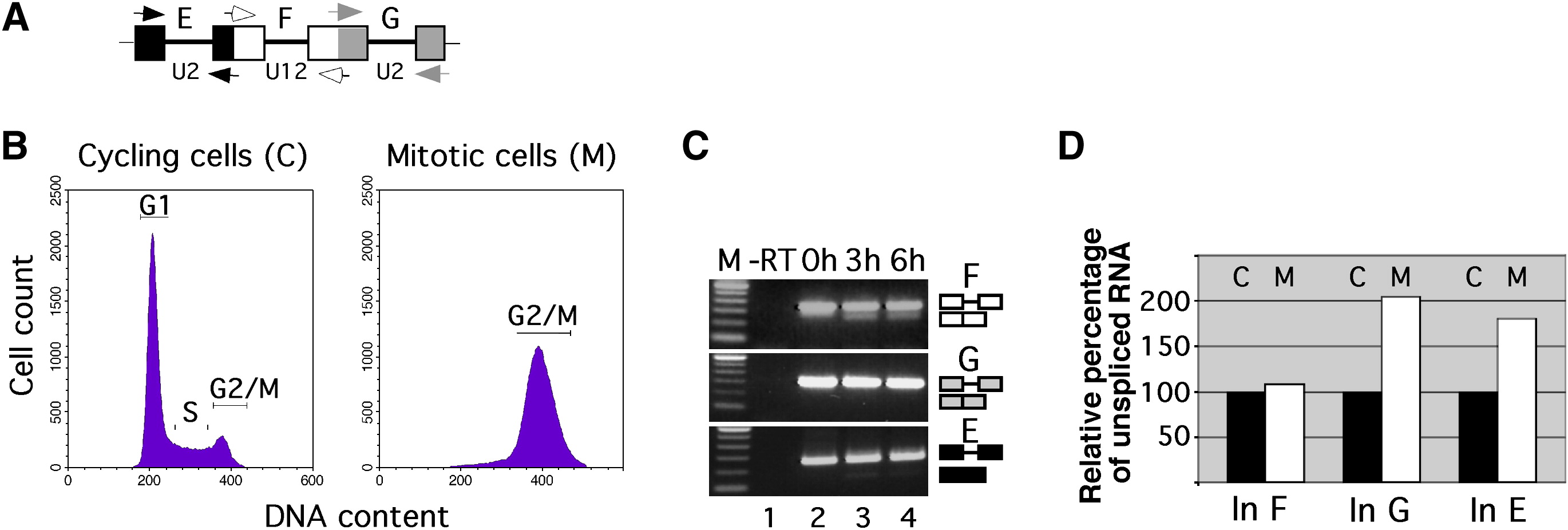
Fig. 3 Minor Splicing Evades Mitotic Downregulation
(A) Scheme of the P120 pre-mRNA region analyzed for splicing (boxes, exons; lines, introns; arrows, primers).
(B) Flow cytometry analysis of cell DNA content from NIH 3T3 cells that grew asynchronously (cycling cells) or were arrested in mitosis by nocodazole treatment.
(C) RT-PCR analysis of U2-type (introns E and G) and U12-type (intron F) splicing of transfected, in-vitro-transcribed P120 minigene pre-mRNA (capped and polyadenylated) in mitotic cells, after the times indicated. For primer positions, see (A). -RT corresponds to RT-PCR reaction lacking reverse transcriptase. Correct splicing was verified by sequencing.
(D) Analysis of splicing of endogenous P120 pre-mRNA in cycling (label C) and mitotically arrested cells (label M). Relative expression levels of mRNA spliced for the major class introns (In) G and E and for the minor-class intron F were measured by real-time quantitative RT-PCR. Values represent the mean of two experiments and are expressed as relative percentage of unspliced RNA (set to 100% in cycling cells).
Reprinted from Cell, 131(4), König, H., Matter, N., Bader, R., Thiele, W., and Müller, F., Splicing Segregation: The Minor Spliceosome Acts outside the Nucleus and Controls Cell Proliferation, 718-729, Copyright (2007) with permission from Elsevier. Full text @ Cell