
Fig. 7
- ID
- ZDB-IMAGE-080829-39
- Publication
- Hsu et al., 2006 - Mosaic Eyes is a novel component of the Crumbs complex and negatively regulates photoreceptor apical size
- All Figures
- Figures for Hsu et al., 2006
|
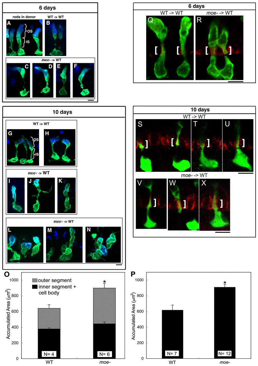
Fig. 7 Moe is required for normal photoreceptor morphology and negatively regulates apical size. (A,B) Wild-type GFP+ rods labeled with anti-Rhodopsin antibody (blue) in a wild-type donor retina (A) and wild-type GFP+ rods transplanted into a wild-type host (B) at 6 dpf. (C-F) moe- GFP+ rods transplanted into wild-type hosts at 6 dpf. (G,H) Wild-type GFP+ rods transplanted into wild-type hosts at 10 dpf. (I-K) moe- GFP+ rods transplanted into wild-type hosts with few moe- neighbors at 10 dpf. (L-N) moe- GFP+ rods transplanted into wild-type hosts with large numbers of moe- neighbors at 10 dpf. (L) Three rods, (M) two to three intermingled rods and (N) three intermingled rods. (O) At 6 dpf, the accumulated area moe- rods is larger than wild-type rods. The increase in size is accounted for largely by an increase in outer segments whereas there is no significanct difference in the inner segments plus cell body (error bars shown; s.e.m.; *, P=0.04). The total accumulated area of moe- rods (901+/-64) is significantly greater than wild-type rods (641+/-37) (P=0.015). Numbers of individuals (N) included. (P) At 10 dpf, the accumulated area of moe- rods is larger than wild-type rods. (*, P=0.001; Student's t-test). (Q-X) panCrb labeling (red) in a single optical section (0.38 μm) of wild-type (Q) and moe- (R) GFP+ rods at 6 dpf and wild-type (S-U) and moe- GFP+ rods (V-X) at 10 dpf. White brackets indicate the region of panCrb localization in the inner segment. Scale bars: 5 μm.