
Fig. S1
- ID
- ZDB-IMAGE-080826-15
- Genes
- Publication
- Mizoguchi et al., 2008 - Sdf1/Cxcr4 signaling controls the dorsal migration of endodermal cells during zebrafish gastrulation
- All Figures
- Figures for Mizoguchi et al., 2008
|
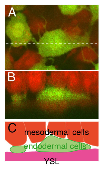
Fig. S1 The endodermal cells lie in the deepest layer of the hypoblast. (A) Confocal image of endodermal and mesodermal cells in Tg(sox17:EGFP) embryo at the 90% epiboly stage. Dextran-Alexa594 was injected into a Tg(sox17:EGFP) embryo at the one-cell stage. The endodermal cells, which have many filopodial processes, are yellowish green and mesodermal cells are red. (B) Reconstructed z-section at the level of the dashed line in A. The extra-embryonic YSL is also red. The endodermal cells occupy the space between the mesodermal cells and extra-embryonic YSL. (C) Schematic representation of B. YSL, yolk syncytial layer.