
|
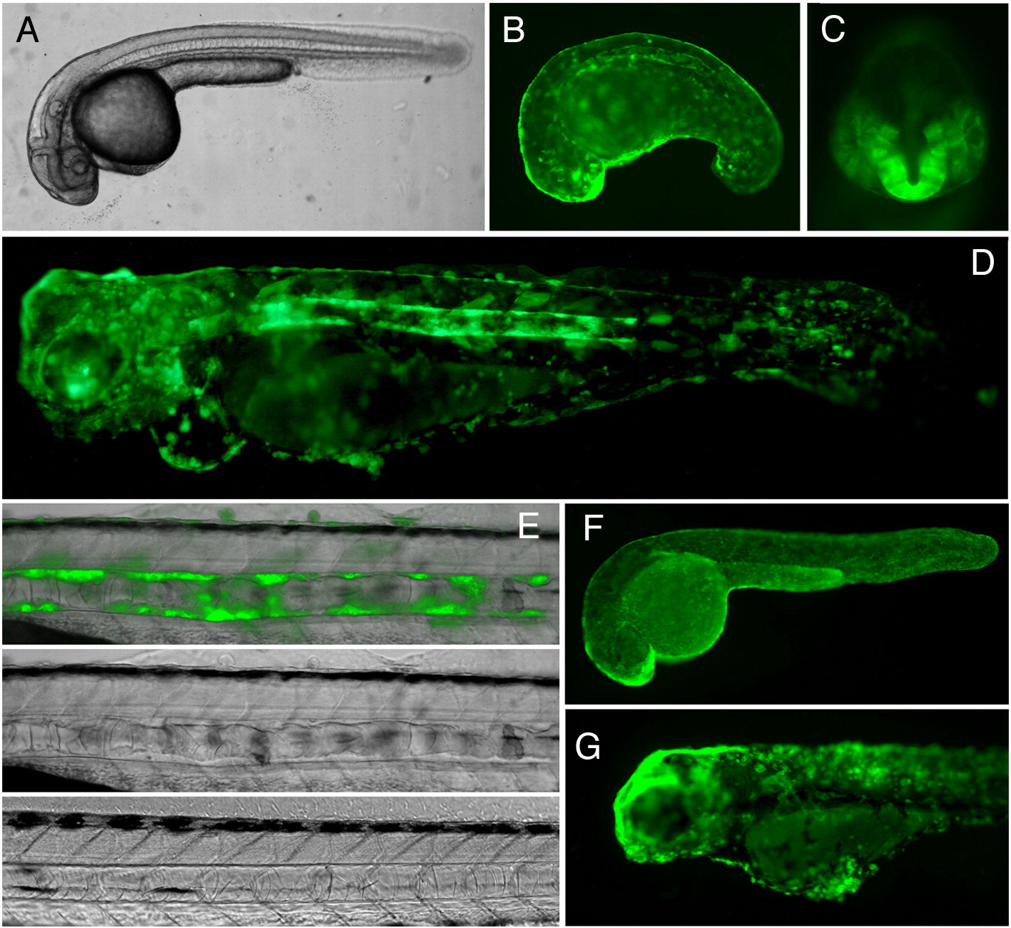
Fig. 5 Inducible expression of EGFP-KrasV12 in transgenic zebrafish. Transgenic zebrafish line harboring insertion of the binary driver construct Ds(krt8:LPR-LOP:EGFP-KrasV12) containing EGFP-KrasV12 under control of the LexOP, F2 (A–E and G). (A) 24 hpf, no induction. (B, C) 24 hpf, 1 μM mifepristone treatment from 10 hpf onwards. GFP fluorescence can be observed in the skin epithelia, the notochord sheath (lateral view, B), in the forebrain (ventral view, C), all of these organs are abnormal. (D) 4 dpf, 1 μM mifepristone treatment 10 hpf onwards, lateral view. Notochord, brain and cranial skeleton are severally affected by the EGFP-KrasV12 expression. Skin epithelial cells lost epithelial cell shape, became globular, formed clusters. (E) Transformation of the sheath cells surrounding the notochord. The top and the middle panel shows the notochord of 4 dpf fish treated with 1 μM mifepristone from 10 hpf onwards. Bottom panel shows notochord of the untreated control. (F) Transgenic control line that harbors the driver-reporter construct pDs(krt8:LPR-LOP:EGFP) containing EGFP gene under control of the LexOP. 24 dpf, 1 μM mifepristone treatment starting at 10 hpf, showing EGFP expression in the skin epithelia. The skin cells have flat epithelial shape and uniformly cover the entire body. (G) 4 dpf. Expression of EGFP-KrasV12 was induced with 1 μM mifepristone treatment from 1 dpf onwards. Due to later induction, there is much less developmental abnormalities, but the cells of skin epithelia cells acquired rounded shape and formed clusters instead of a continuous layer.
Reprinted from Developmental Biology, 320(1), Emelyanov, A., and Parinov, S., Mifepristone-inducible LexPR system to drive and control gene expression in transgenic zebrafish, 113-121, Copyright (2008) with permission from Elsevier. Full text @ Dev. Biol.