
|
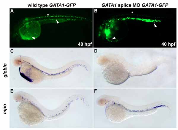
Fig. S2 Wild-type gata1-egfp embryos have GFP in circulating blood cells in the vessels and over the yolk (A, arrowheads) and ectopically in the neural tube (asterisk). A gata1 morpholino targeting the exon/intron splice sites was used to avoid interference with transgene expression. At 48 hpf, Gata1-deficicent embryos had large, round, noncirculating GFP-positive cells distributed in a pattern resembling mpo expression (B). gata1 MO-injected gata1-egfp embryos express gfp in noncirculating cells in the major vessels and on the yolk (B, arrowheads). be1 globin (C) and mpo (E) are expressed normally in wild-type embryos. In gata1 MO-injected embryos, be1 globin expression is absent (D) and mpo expression is expanded (F).
Reprinted from Developmental Cell, 8(1), Galloway, J.L., Wingert, R.A., Thisse, C., Thisse, B., and Zon, L.I., Loss of gata1 but not gata2 converts erythropoiesis to myelopoiesis in zebrafish embryos, 109-116, Copyright (2005) with permission from Elsevier. Full text @ Dev. Cell