
Fig. 9
- ID
- ZDB-IMAGE-080729-8
- Antibodies
- Publication
- Craig et al., 2008 - Expression and Regulation of the Vitamin D Receptor in the Zebrafish, Danio rerio
- All Figures
- Figures for Craig et al., 2008
|
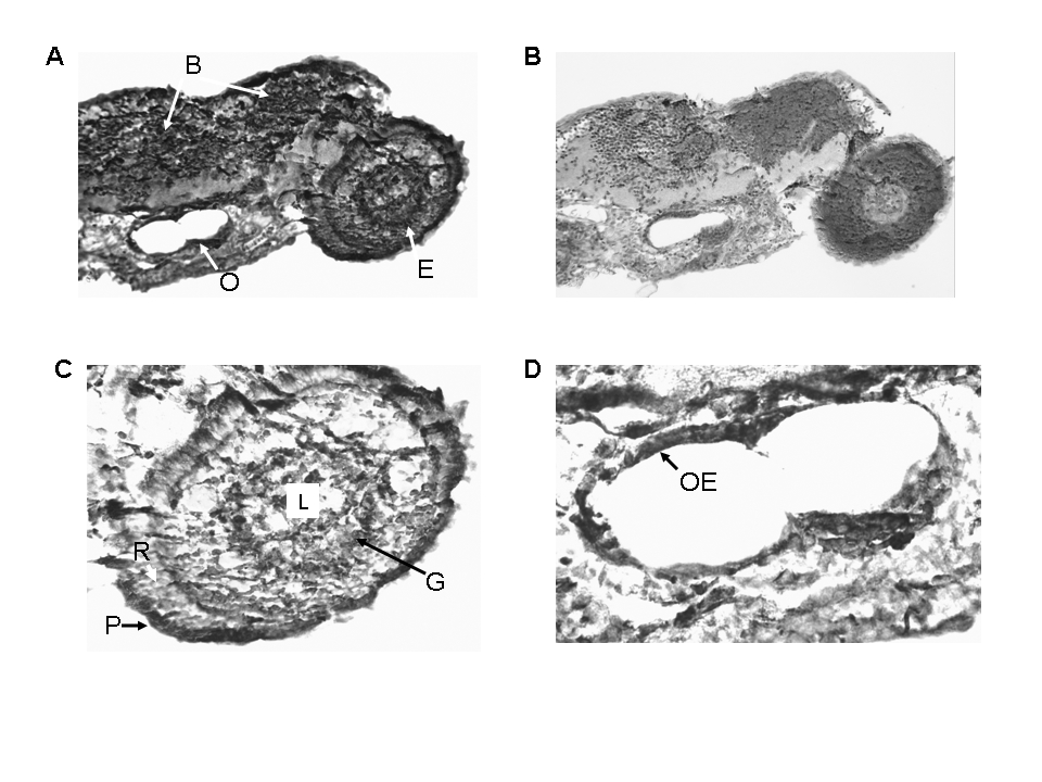
Fig. 9 Immunostaining of 96 hour post-fertilization zebrafish embryo with anti-VDR antibody or pre-immune serum. 100 X original magnification. Panel A. 96 hour post-fertilization embryo immunostained with anti-VDR antibody. Note staining (brown color) of cells within the eye (E), the brain (B) and the otic vesicle (O). 100 X original magnification. Panel B. 96 hour post-fertilization embryo immunostained with pre-immune serum. Note absence of staining for the VDR. 100 X original magnification. Panel C. Section of the eye immunostained with anti-VDR antibody; 200 X original magnification. L = lens; G = ganglion cells; R = photoreceptor cells; P = pigmented epithelial cells. Panel D. Otic vesicle immunostained with anti-VDR antibody. 200 X. original magnification.