
Fig. 6
- ID
- ZDB-IMAGE-080729-10
- Antibodies
- Publication
- Craig et al., 2008 - Expression and Regulation of the Vitamin D Receptor in the Zebrafish, Danio rerio
- All Figures
- Figures for Craig et al., 2008
|
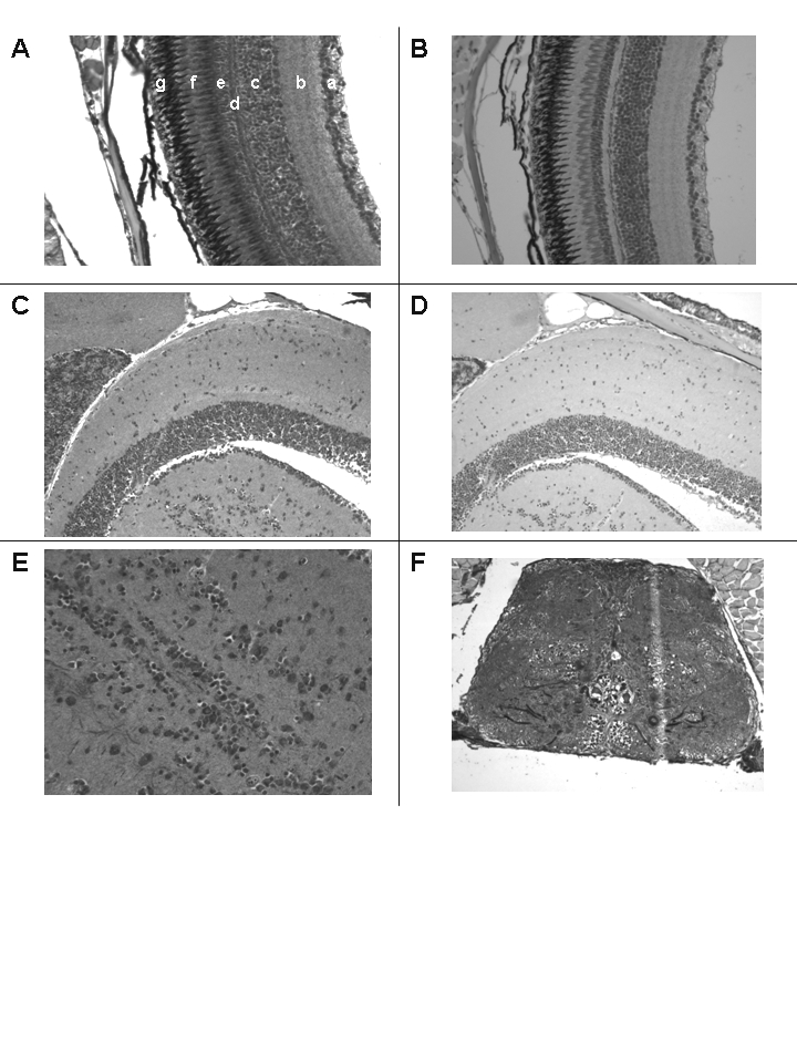
Fig. 6 Immunostaining of the eye and neural tissues with anti-VDR antibody or pre-immune serum. 200 X original magnification. Panel A. Section of the retina immunostained with anti-VDR antibody. a = ganglion cell layer; b = inner plexiform layer; c = inner nuclear layer; d = outer plexiform layer; e = outer nuclear layer; f = rods; g = pigmented layer. Panel B. Section of retina using pre-immune serum. Note absence of staining in all layers. Panel C. Section of brain immunostained with anti-VDR antibody. Panel D. Section of brain immunostained with pre-immune serum. Panel E. Section of brain immunostained with anti-VDR antibody (400 X). Panel F. Section of spinal cord staining with anti-VDR antibody.