
Fig. 4
- ID
- ZDB-IMAGE-080715-28
- Genes
- Publication
- Liu et al., 2008 - In vivo time-lapse imaging delineates the zebrafish pituitary proopiomelanocortin lineage boundary regulated by FGF3 signal
- All Figures
- Figures for Liu et al., 2008
|
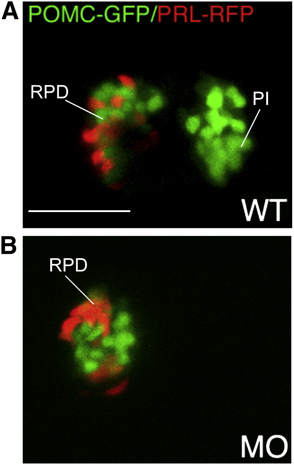
Fig. 4 Lineage specific suppression of PI POMC cells in FGF3 morphants. 1–2 cell stage POMC-GFP/PRL-RFP double transgenic embryos were either injected with 0.4 ng of FGF3 MO (B) or vehicle (A) and then cultured in routine medium. At 56 hpf, pituitary POMC-GFP/PRL-RFP expression patterns were analyzed in live embryos using confocal microscopy. In FGF3 morphant, POMC-GFP expressing cells are detected within the rostral pars distalis but not in the pars intermedia, whereas PRL-RFP expression is intact. Ventral pituitary view under high magnification (63x), anterior to the left. WT, wild type; MO, FGF3 morphant. RPD, rostral pars distalis; PI, pars intermedia. Scale bar, 50 μm.
Reprinted from Developmental Biology, 319(2), Liu, N.A., Ren, M., Song, J., Ríos, Y., Wawrowsky, K., Ben-Shlomo, A., Lin, S., and Melmed, S., In vivo time-lapse imaging delineates the zebrafish pituitary proopiomelanocortin lineage boundary regulated by FGF3 signal, 192-200, Copyright (2008) with permission from Elsevier. Full text @ Dev. Biol.