
|
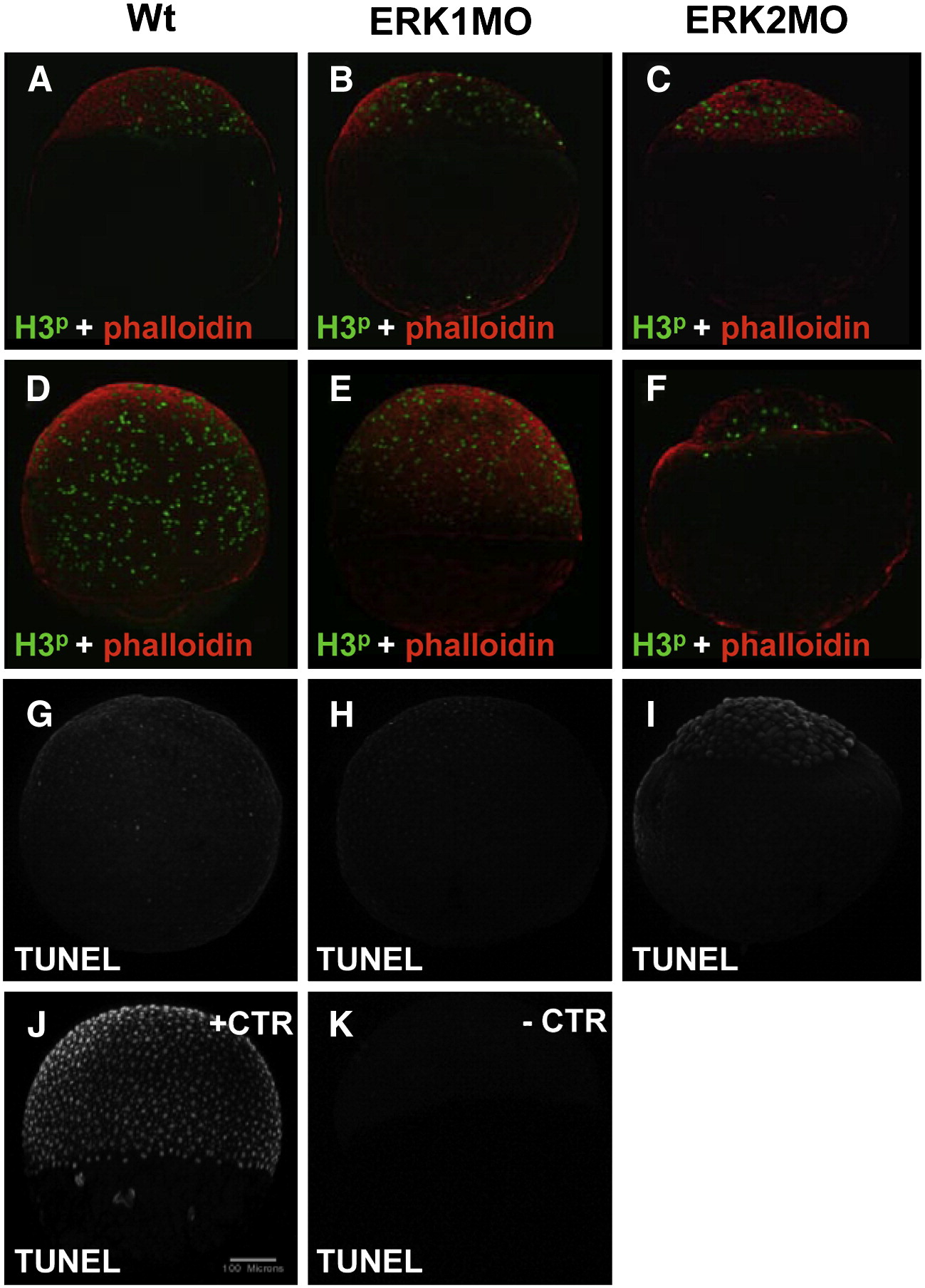
Fig. S5 Depletion of ERKs does not induce proliferation or apoptosis defects during early developmental stages. Proliferation was analyzed by immuno-histochemistry (A–F) of phospho-histone H3 (H3p) and counterstained with Phalloidin-rhodamin of wild type ERK1MO and ERK2-O injected embryos at 4.5 (A–C) and 8 hpf (D–F). TUNEL in situ cell-death staining in wild type embryos (G) compared to ERK1MO (H) and ERK2MO (I) injected embryos at 8 hpf (G–K). As a positive control, wild type embryos were treated with DNAse after fixation (J; + CTR), which resulted in positive DNA staining of the nuclei. As a negative control, embryos were stained with developer only (K; - CTR), which gave no background signal.
Reprinted from Developmental Biology, 319(2), Krens, S.F., He, S., Lamers, G.E., Meijer, A.H., Bakkers, J., Schmidt, T., Spaink, H.P., and Snaar-Jagalska, B.E., Distinct functions for ERK1 and ERK2 in cell migration processes during zebrafish gastrulation, 370-383, Copyright (2008) with permission from Elsevier. Full text @ Dev. Biol.