
|
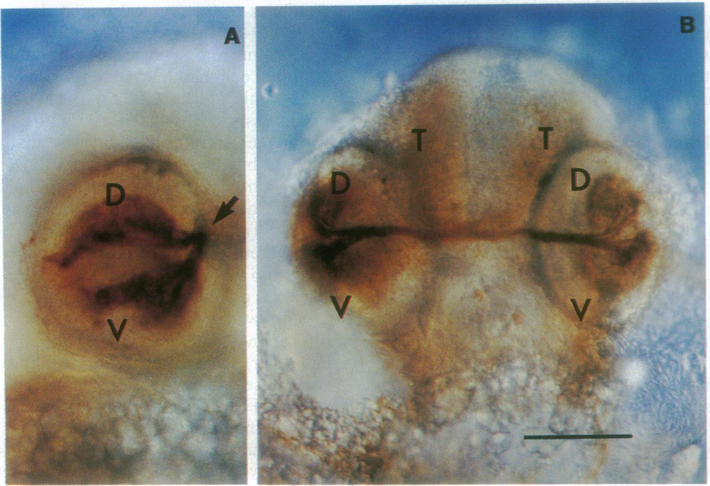
Fig. 3 Ganglion cells and their axons stained with a monoclonal antibody in retinoic acid-treated animals. The embryos were treated with, 1 μM retinoic acid for 2 hr at the 2- to 3-somite stage and raised in 0.2 mM phenylthiocarbamide to suppress pigment formation. At 3.5 days of development, the embryo was fixed and processed for whole-mount staining. (A) Two fields (D and V) of ganglion cells can be seen in this retinoic acid-treated eye viewed with Nomarski optics from the side of the embryo. Bundles of axons project from each ganglion cell field and join to project centrally (arrow). (B) A view from the front of the head showing how the bundles of axons join as they exit the eye and innervate the contralateral tecta (T). (Bar = 170 μm.)