
|
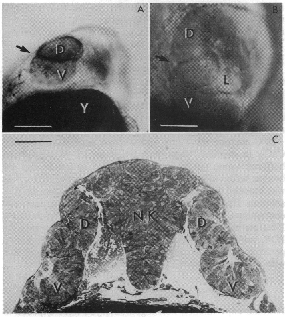
Fig. 1 (A) Bifurcated eye in a living zebrafish embryo viewed with a dissecting microscope. The bifurcation (arrow) divides the eye into dorsal (D) and ventral (V) regions. In this case, pigment cells line the bifurcation. The embryo was treated 10.5 hr after fertilization with 1 μM retinoic acid for 1 hr and viewed 15 hr later. Y, yolk. (Bar = 140 μm.) (B) Bifurcated eye viewed by Nomarski optics in a living zebrafish embryo. Two retinas are readily visualized; one is located dorsally (D) to the bifurcation (arrow); the other is positioned ventrally (V). A lens (L) is associated with the ventral retina. The embryo in this case was treated at a late gastrula stage with 1 μM retinoic acid for 1 hr and raised, thereafter, in 0.2 mM phenylthiocarbamide to suppress pigment formation. The photomicrograph was taken 30 hr after fertilization. (Bar = 95 μm.) (C) Conventional light micrograph of a section through the head region of a zebrafish embryo. Dorsal (D) and ventral (V) retinas are present on both sides of the neural keel (NK). (Bar = 120 μm.)