
|
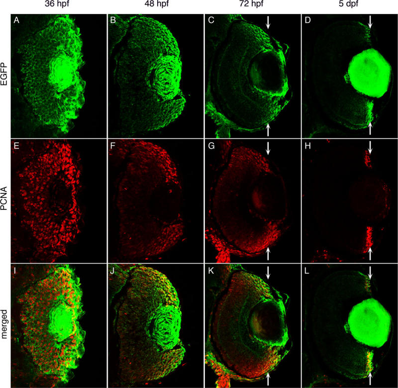
Fig. 3 Enhanced green fluorescent protein expression during retinal development in Tg(ccnb1:EGFP)nt18 zebrafish. Enhanced green fluorescent protein (EGFP; A-D), proliferating cell nuclear antigen (PCNA; E-H), and merged expression patterns (I-L) are shown at 36, 48, 72 hpf, and 5 dpf of retinal development. At 36 hpf, PCNA and EGFP expression are almost ubiquitous throughout the retina (A, E, I). At 48 hpf, EGFP and PCNA expression is limited in the central retina, but persists near the retinal margin (B, F, J). At 72 hpf, EGFP and PCNA expression are largely absent in the central retina and become further focused in the cells near the margin (C, G, K; arrows). By 5 dpf, EGFP and PCNA expression are restricted to the retinal margin (D, H, L; arrows). At all time points, EGFP expression but not PCNA persists in the lens.