
Fig. 5
- ID
- ZDB-IMAGE-080604-41
- Antibodies
- Publication
- Chi et al., 2008 - Genetic and Physiologic Dissection of the Vertebrate Cardiac Conduction System
- All Figures
- Figures for Chi et al., 2008
|
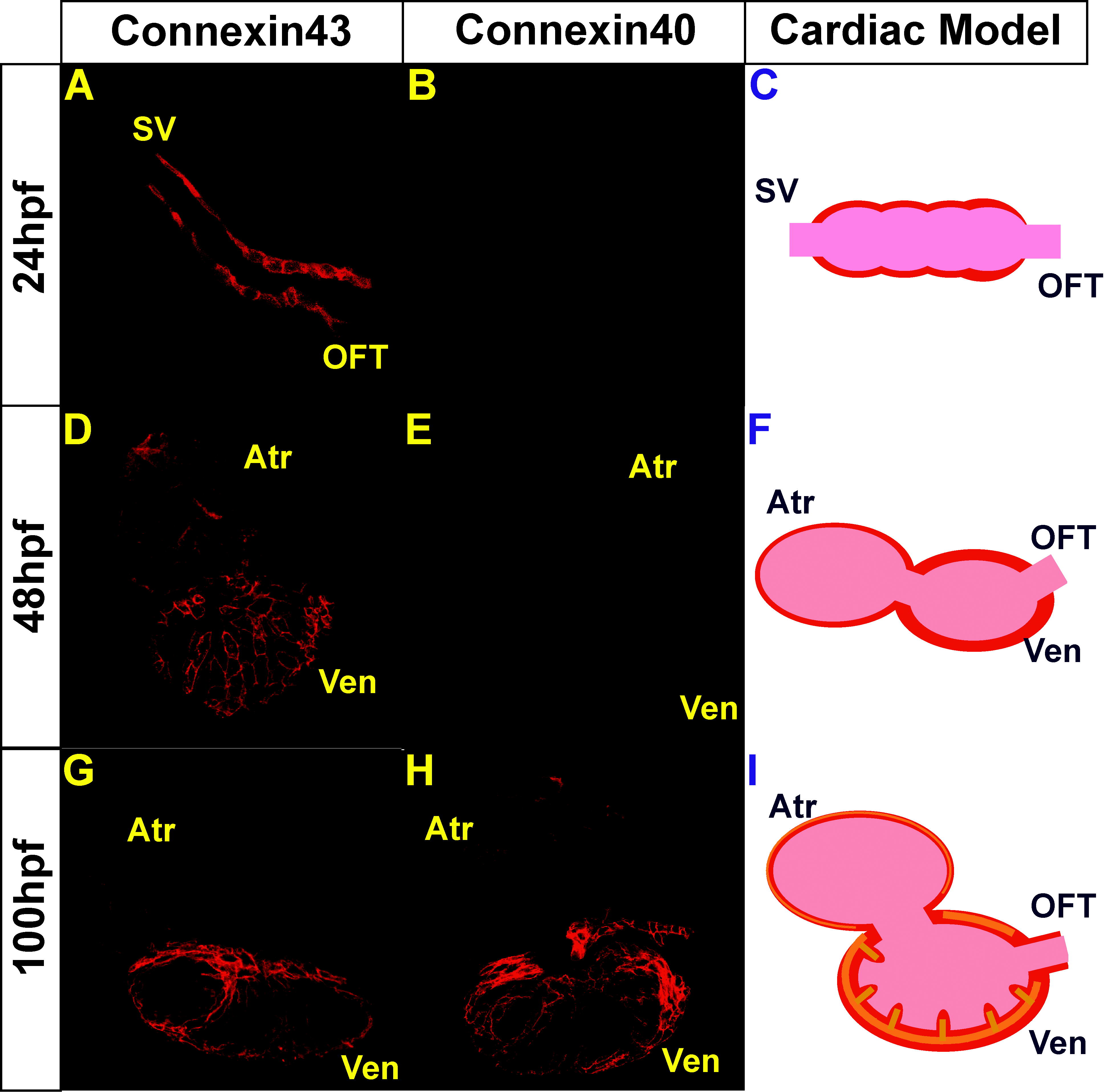
Fig. 5
Immunolocalization of Cx40 and Cx43 in Wild-Type Hearts
Confocal images of the heart at 24, 48, and 100 hpf, following immunostaining with anti-Cx40 or anti-Cx43 antibodies (red).
(A, D, and G) Cx43 immunoreactivity was observed throughout the myocardium at 24, 48, and 100 hpf while (B, E and H) Cx40 immunoreactivity was observed weakly at 48 hpf and strongly at 100 hpf.
(C, F and I) Schematic representation of Connexin staining at each developmental stage. Cx43 immunoreactivity is observed throughout the myocardium at all stages and represented in red, and Cx40 immunoreactivity is observed at later cardiac developmental stages and represented in orange.