
Fig. 3
- ID
- ZDB-IMAGE-080604-39
- Genes
- Publication
- Chi et al., 2008 - Genetic and Physiologic Dissection of the Vertebrate Cardiac Conduction System
- All Figures
- Figures for Chi et al., 2008
|
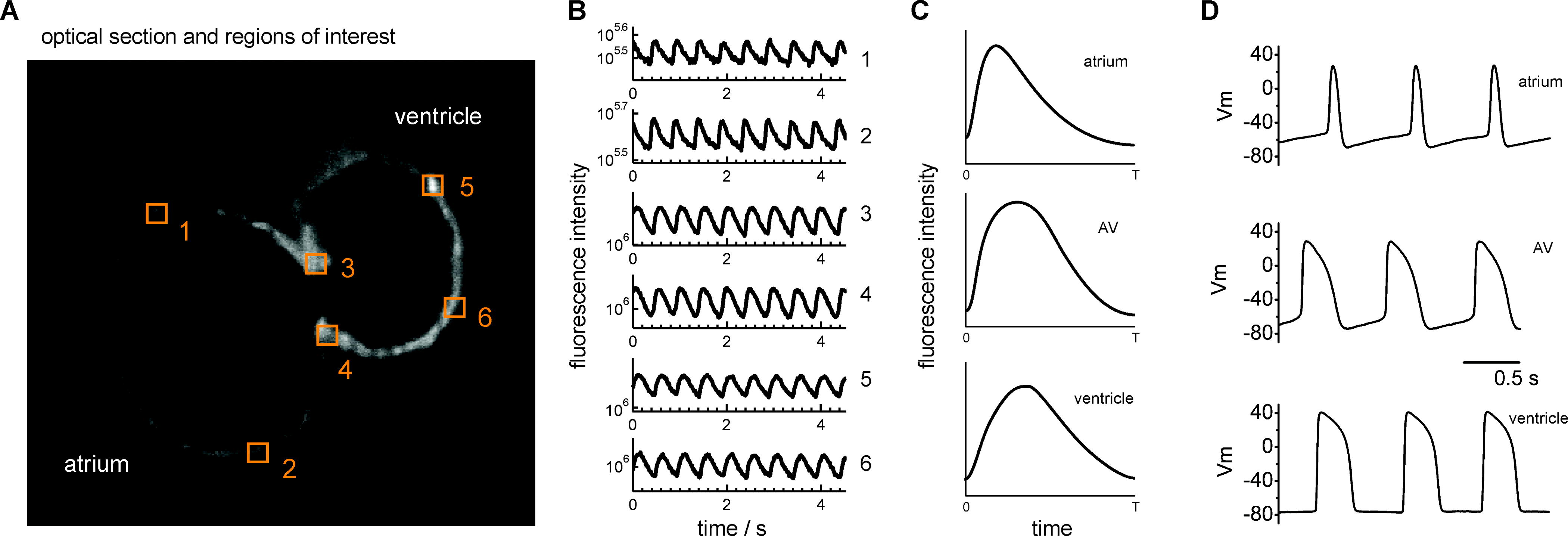
Fig. 3
Calcium Transients and APs Confirm That AV Myocardium Has Distinct Electrophysiologic Properties That Distinguish It from Atrium and Ventricle
(A) Optical section of 48 hpf Tg(cmlc2:gCaMP)s878 heart. Numbers represent areas where calcium transients for the atrium, ventricle, and AV canal were recorded.
(B) Fluorescence intensity of a single pixel from each region was recorded to obtain calcium transients and plotted over time in seconds. All plots are semilogarithmic and identically scaled.
(C) Average calcium transient for each cardiac region.
(D) The APs from each of these regions were recorded in wild-type explanted 48 hpf hearts using patch pipettes and current clamp techniques. Distinct calcium transients and APs were detected in atrium, AV canal, and ventricle.