
Fig. 1
- ID
- ZDB-IMAGE-080604-1
- Antibodies
- Publication
- Postel et al., 2008 - Zebrafish integrin-linked kinase is required in skeletal muscles for strengthening the integrin-ECM adhesion complex
- All Figures
- Figures for Postel et al., 2008
|
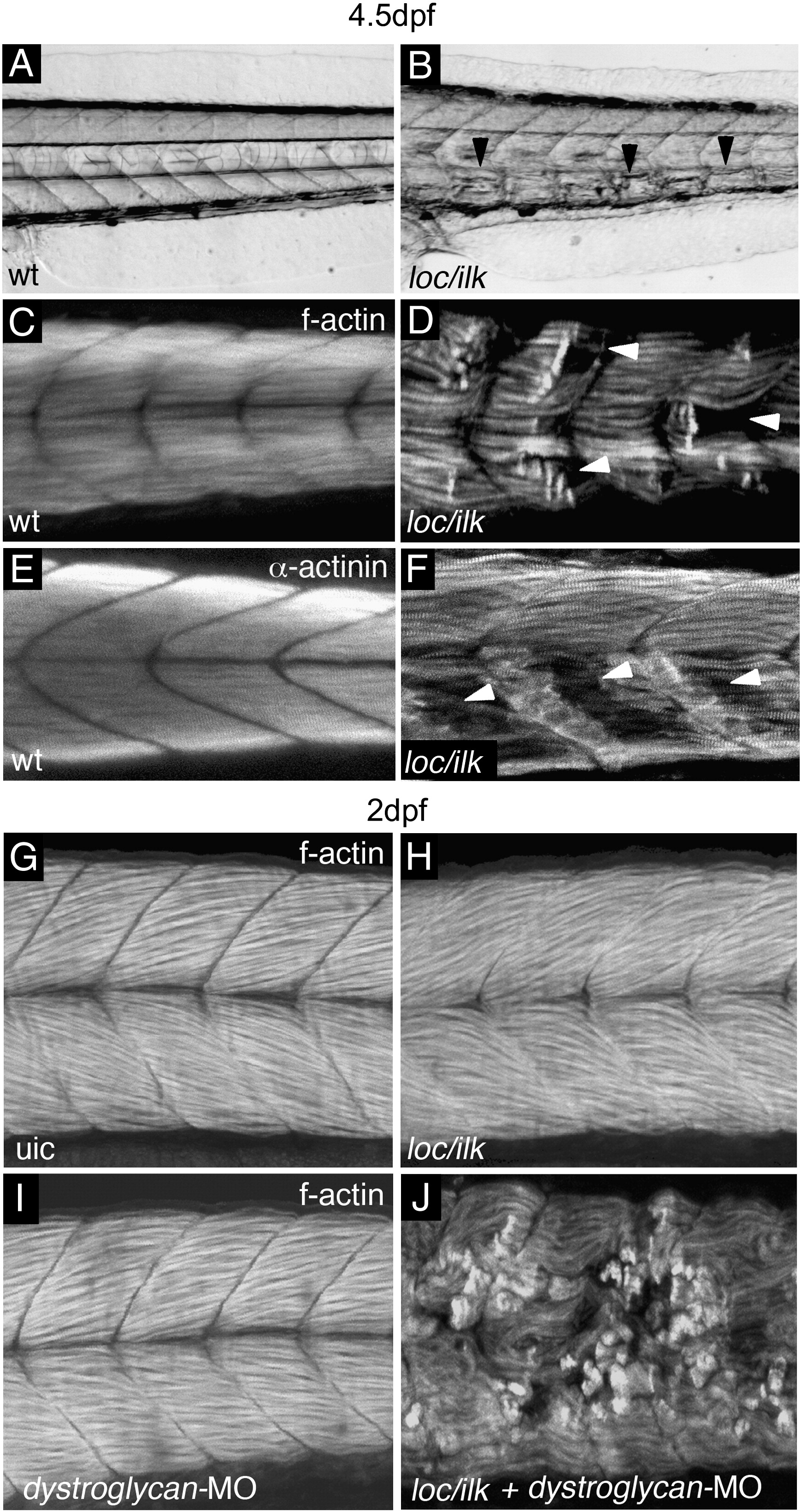
Fig. 1 Skeletal muscle detachments in loc/ilk mutant embryos. (A,B) Transmitted light images of tail regions in wt (A) and loc/ilk mutant embryos (B) at 4.5 dpf. Muscle fibre retractions are indicated with arrowheads. (C,D) Phalloidin staining and confocal images of wt sibling embryo (C) and of loc/ilk mutant embryo (D) at 4.5 dpf. Muscle fibre detachments are apparent in 50% of the loc/ilk mutant embryos (arrowheads). (E,F) α-actinin antibody staining of wt sibling (E) and loc/ilk mutant embryo (F) at 4.5 dpf. Regions of muscle fibre detachments are indicated by arrowheads. (G–J) Phalloidin staining and confocal images of uninjected control embryo (G), uninjected loc/ilk mutant embryo (H), dystroglycan MO injected wt embryo (I) and a dystroglycan MO injected loc/ilk mutant embryo (J). Tail regions of 2 dpf embryos are shown.
Reprinted from Developmental Biology, 318(1), Postel, R., Vakeel, P., Topczewski, J., Knöll, R., and Bakkers, J., Zebrafish integrin-linked kinase is required in skeletal muscles for strengthening the integrin-ECM adhesion complex, 92-101, Copyright (2008) with permission from Elsevier. Full text @ Dev. Biol.