
|
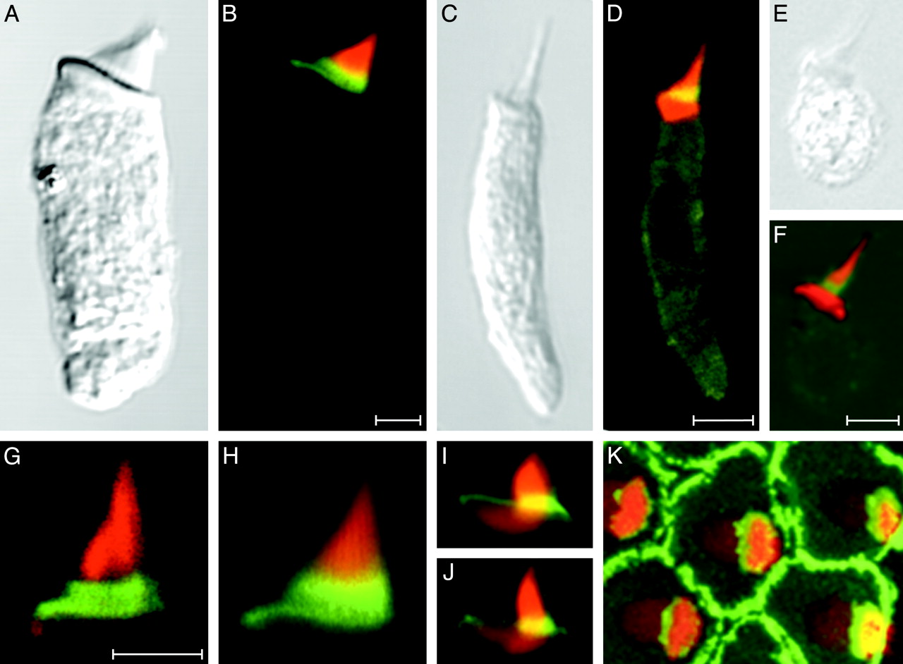
Fig. 3 Anti-radixin immunolabeling of hair bundles from various vertebrate classes. (A and B) A hair cell isolated from the bullfrog's sacculus displays strong anti-radixin labeling (green) at the base of the hair bundle; the yellow signal results from partial overlap of this labeling with that by fluorescent phalloidin (red). (C and D) A hair cell from the lagena of the zebrafish exhibits anti-radixin labeling at the hair bundle's base. (E and F) Anti-radixin immunolabeling occurs at the hair bundle's base in a hair cell isolated from the utriculus of a mouse. (G and H) Higher-magnification views of two hair bundles from the frog's sacculus demonstrate the most intense anti-radixin labeling at the stereociliary bases. (I and J) In higher-magnification views of hair bundles from short hair cells of the chicken's cochlea, anti-radixin labeling is again strongest at the stereociliary bases. The complex shape of the cuticular plate is also apparent. (K) A surface view of a cluster of short hair cells isolated from the chicken's cochlea demonstrates intense anti-radixin labeling of the supporting cells surrounding each hair cell. An additional immunofluorescence signal is evident at the bases of the stereocilia, which are seen in a stack of 73 images acquired at 0.18-μm intervals. All preparations have additionally been labeled with phalloidin conjugated to Alexa Fluor 568 (red). (Scale bars, 5 μm; bar in B applies also to A; bar in D applies also to C, I, J, and K; bar in F applies also to E; bar in G applies also to H.)