
Fig. 3
- ID
- ZDB-IMAGE-080529-3
- Publication
- Sapede et al., 2005 - Role of SDF1 chemokine in the development of lateral line efferent and facial motor neurons
- All Figures
- Figures for Sapede et al., 2005
|
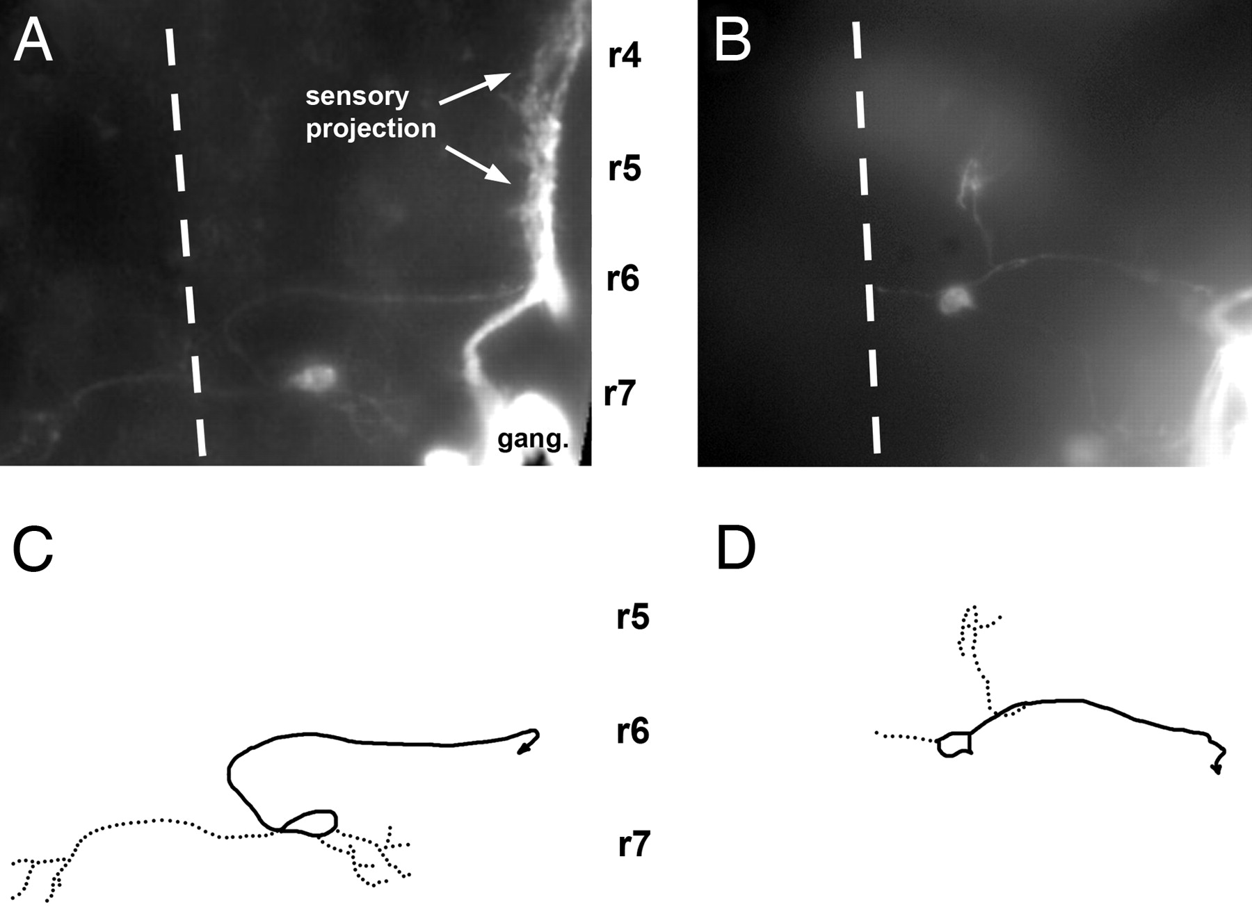
Fig. 3 Central pathway of caudal PLL efferent neurons in 3-day-old embryos. (A and C) In the wild type. (B and D) In a sdf1a morphant embryo. A and B have been assembled by using a set of photographs taken at various focal planes; C and D have been drawn on the basis of A and B. Solid lines, axons; dotted lines, dendrites; dashed line, midline; r4, r5, r6, and r7, rhombomeres 4, 5, 6, and 7. In this and in the following figures, the approximate positions of rhombomeres shown on the figures are based on two criteria: the position of the otic vesicle, which forms along rhombomere 5 and extends approximately from rhombomere 4 to rhombomere 6, and the position of identified cranial nerves, specifically the V and VII nerves that exit the brain at the levels of rhombomeres 2 and 4, respectively, and were visualized by fluorescence in islet-GFP embryos, both in wild type and inmorphants.